
Drawing Of Foot Bones
Drawing Of Foot Bones - Since the foot is so bony, knowing the inside anatomy directly helps you draw the outside surface. Web all 26 bones of the foot are described generally for drawing purposes. The form of the foot is considerably made of the form of the bones. Web your assignment is to simplify the foot bones into their basic forms. Human foot bone foot bone structure foot bone diagram foot bone vector sort by:
Web hindfoot talus calcaneus the talus connects the foot to the rest of the leg and body through articulations with the tibia and fibula, the two long bones in the lower leg. A simple way to draw feet is to begin by drawing the sole of the foot. Web fortunately, the bones are a great way to study the foot. Pinch in/out or mousewheel or ctrl + left mouse button Web learn the bones of the foot in half the time with these interactive quizzes and labeling activities! Web there are 26 bones in the foot. The phalanges, which are the bones in your toes.

Foot & Ankle Bones
Web if you missed the foot bone lesson, go watch it! Web there are 26 bones in the foot. A simple way to draw feet is to begin by drawing the sole of the foot. Bones, muscles, ligaments, and tendons make up the foot. Let’s look briefly at the structure of the foot: The skeletal.
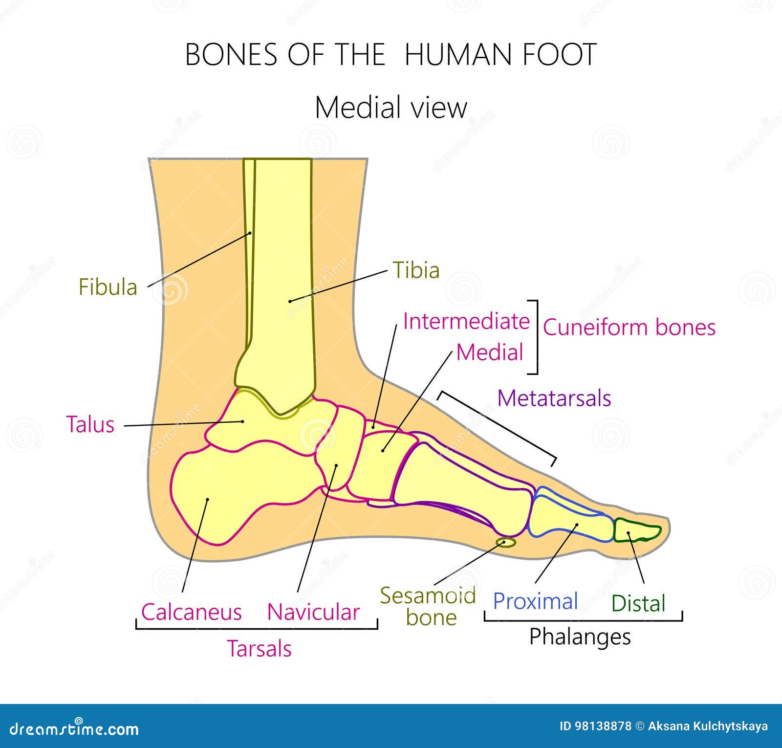
Anatomy_bones of the Human Foot Medial View Stock Vector Illustration
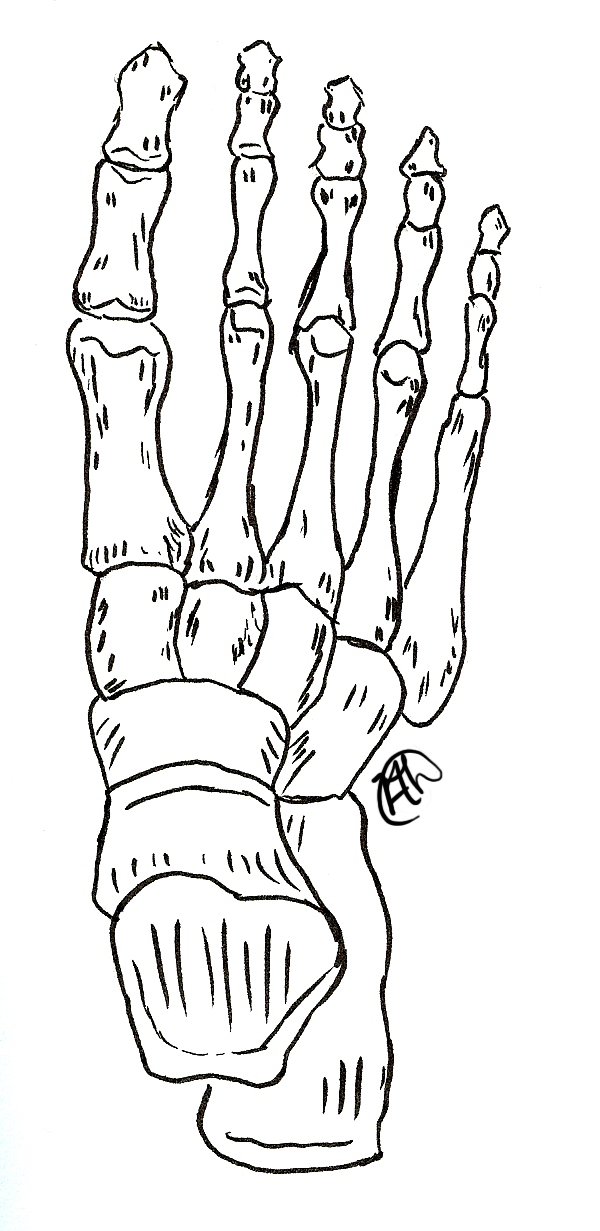
Most popular bones of the foot and ankle joint medical vector illustration. When you know the bones, you're 90% ready to draw a foot. The cuneiform bones, the navicularis, and the cuboid, all of which function to give your foot a solid yet somewhat flexible foundation. Web hindfoot talus calcaneus the talus connects the foot.
Skeleton Feet Drawing at Explore collection of
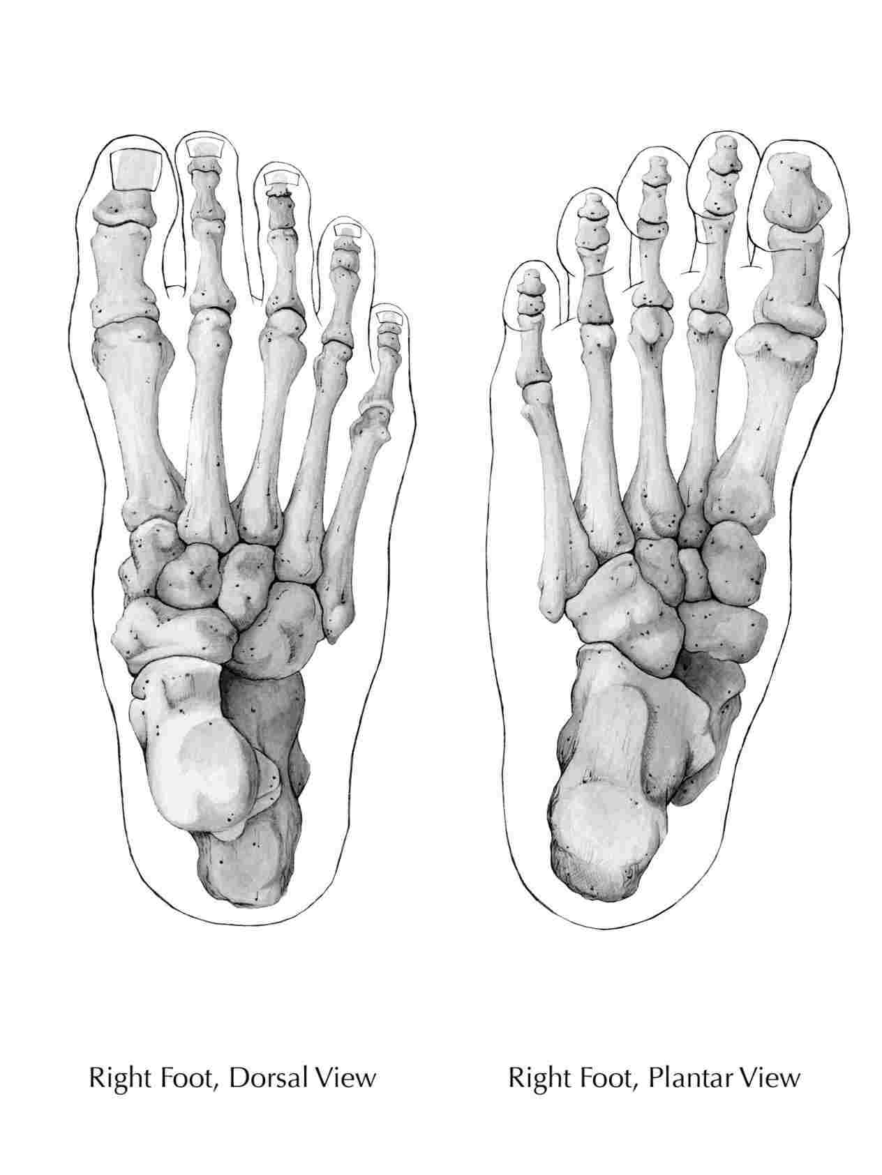
The bones of the foot. The distal tarsals are the cuboid and three cuneiform bones (lateral, intermediate, and medial). No toes, no arches, just the. How to draw feet basics of the foot. Midfoot navicular cuboid medial cuneiform intermediate cuneiform lateral cuneiform The 3d model of the robo foot. The metatarsals, which run through the.
.jpg)
Foot Bone Diagram resource Imageshare
Web the talus, or ankle bone: Web hindfoot talus calcaneus the talus connects the foot to the rest of the leg and body through articulations with the tibia and fibula, the two long bones in the lower leg. Side view of skeleton leg with phalange, metatarsal, tarsal and calcaneus, cuneiform, navicular and tibia bones diagram..
.jpg)
Foot Bone Diagram resource Imageshare
Side view of skeleton leg with phalange, metatarsal, tarsal and calcaneus, cuneiform, navicular and tibia bones diagram. This cornerstone is not altered as in brick work, but rather moves openly between the inward also, external condyle. The cuneiform bones, the navicularis, and the cuboid, all of which function to give your foot a solid yet.
Bones of the Feet ClipArt ETC
Web the 26 bones of the foot consist of eight distinct types, including the tarsals, metatarsals, phalanges, cuneiforms, talus, navicular, and cuboid bones. The bone structure of the foot consists of three different sections: No toes, no arches, just the. This lesson will focus on the overall design of the foot, and the form, proportion,.

Bones of the Foot Anatomy Sketch
Since the foot is so bony, knowing the inside anatomy directly helps you draw the outside surface. Human foot bone foot bone structure foot bone diagram foot bone vector sort by: Bones, muscles, ligaments, and tendons make up the foot. Midfoot navicular cuboid medial cuneiform intermediate cuneiform lateral cuneiform Web orbit navigation move camera: Most.
Foot Skeleton Drawing at GetDrawings Free download
The bones of the foot are divided into anterior region, posterior region, dorsal region, plantar region, distal region, proximal region, medial region, and lateral region. This cornerstone is not altered as in brick work, but rather moves openly between the inward also, external condyle. Web your assignment is to simplify the foot bones into their.

Foot Bone Anatomy Vector Illustration 539973 Vector Art at Vecteezy
The 3d model of the robo foot. The bones of the foot are divided into anterior region, posterior region, dorsal region, plantar region, distal region, proximal region, medial region, and lateral region. When you know the bones, you're 90% ready to draw a foot. No toes, no arches, just the. The bones of the foot..

Skeleton Foot by Isasan on DeviantArt
Bones, muscles, ligaments, and tendons make up the foot. Most popular bones of the foot and ankle joint medical vector illustration. Pinch in/out or mousewheel or ctrl + left mouse button Web the talus, or ankle bone: Web this article includes a diagram showing the bones of the foot, which will give an insight about.
Drawing Of Foot Bones Most popular human foot bones front and side view anatomy The calcaneus, or heel bone : Since the foot is so bony, knowing the inside anatomy directly helps you draw the outside surface. Foot bones vector sketch of human anatomy, orthopedics medicine design. Most popular bones of the foot and ankle joint medical vector illustration.
The 3D Model Of The Robo Foot.
When you know the bones, you're 90% ready to draw a foot. The skeletal structure of the foot. The phalanges, which are the bones in your toes. Web there are 26 bones in the foot.
Web The Talus, Or Ankle Bone:
This cornerstone is not altered as in brick work, but rather moves openly between the inward also, external condyle. The proximal tarsal bones are the talus and calcaneus. Web the bones of the foot are wedged together and bound by ligaments. You'll find still images of foot bone poses in the download below.
The Tarsals Or Ankle Bones In Blue, The Metatarsi.
Side view of skeleton leg with phalange, metatarsal, tarsal and calcaneus, cuneiform, navicular and tibia bones diagram. Web an anatomy drawing and text of the skeleton of the foot, from the 19th century. Most popular human foot bones front and side view anatomy Web this article includes a diagram showing the bones of the foot, which will give an insight about them.
A Simple Way To Draw Feet Is To Begin By Drawing The Sole Of The Foot.
Anatomy of the human body torso (163 lessons) 0% completed arms (101 lessons) 0% completed legs (107 lessons) 0% completed leg bones foot bones butt muscles inner leg muscles quadriceps. The form of the foot is considerably made of the form of the bones. This lesson will focus on the overall design of the foot, and the form, proportion, and mobility of the individual bones. Most popular bones of the foot and ankle joint medical vector illustration.