
The Drawing And Photomicrograph
The Drawing And Photomicrograph - It has been long argued that students can be weak in perceiving microscopic entities compared to macroscopic entities. At a basic level, photomicroscopy may be performed simply by connecting a camera to a microscope, thereby enabling the user to take photographs at reasonably high magnification. For example, grain supported structures and fractured grains are common in many sandstones ( fig. Web arteries, veins & capillaries: The drawing and photomicrograph below show a relaxed sarcomere.
Web the drawing and photomicrograph given shows a relaxed sarcomere. The walls of arteries and veins contain the same components; A actin filament b a band c h zone d i band (2) h e. The drawing and photomicrograph given shows a relaxed sarcomere. The drawing and photomicrograph below show a relaxed sarcomere. The drawing and photomicrograph below show a relaxed sarcomere. Web a light micrograph or photomicrograph is a micrograph prepared using an optical microscope, a process referred to as photomicroscopy.
Solved s. The drawing and photomicrograph below show a
In the photomicrograph below of compact bone tissue, find and label the indicated structures. Using the terms from the key, identity each structure indi cated by a leader ine or bracket. Each bundle, in turn, is composed of many large peripheral axons. The drawing and photomicrograph below show a relaxed sarcomere. The number 2 in.

Photomicrographs and annotated sketches of microstructural textures in
The number of pixels determines how big a digital image can be before it looks “pixilated”. Web the drawing and photomicrograph given shows a relaxed sarcomere. Web the objective of our study was to establish a detailed photomicrographing protocol for pathologists and dermatopathologists using standard overhead camera and image editing packages. Obtain a slide of.

Photomicrograph (a; as seen from dorsal) and schematic drawing (b) of
The walls of the capillaries are formed from a. The drawing and photomicrograph below show a relaxed sarcomere using the rhe we beldce cated by a leader line or bracket. Fill out the blanks next to your drawing. A actin filament b a band c h zone d i band (2) h e. The number.
Solved 5. The drawing and photomicrograph below show a
Web the drawing and photomicrograph below show a relaxed sarcomere. Web the objective of our study was to establish a detailed photomicrographing protocol for pathologists and dermatopathologists using standard overhead camera and image editing packages. 11.2 ), are remarkably similar to chondrules, the main component of chondrites ( fig. The drawing and photomicrograph below show.
Solved 5. The drawing and photomicrograph below show a
Z disc (2) solution verified answered 6 months ago create a free account to view solutions View the slide on an appropriate objective. The number 2 i parentheses indicates that the structure will be labeled twice. But in differing proportions and with different wall thicknesses. The drawing and photomicrograph given shows a relaxed sarcomere. The.
Solved s. The drawing and photomicrograph below show a
A actin filament b a band c h zone d i band (2) h e. A band c, h zone d. A columnar epithelial cell looks like a column or a tall rectangle. The walls of the capillaries are formed from a. The number of pixels, the dynamic range (maximum number of electrons per pixel),.

A and B A light microscopic photomicrograph and drawing of young ♀ C
Details of the structures inside the goblet cell can be seen in an electron micrograph. Drawing making biological drawings (for teachers) new senior secondary mastering biology 4 The drawing and photomicrograph below show a relaxed sarcomere. Web there are three basic shapes used to classify epithelial cells. At a basic level, photomicroscopy may be performed.

Photomicrographs and drawings of selected strains. 10 m m
A actin filament b a band c h zone d i band (2) h e. Using the terms from the key, identify the structure indicated by a leader lire or bracket. 11.2 ), are remarkably similar to chondrules, the main component of chondrites ( fig. Web a light micrograph or photomicrograph is a micrograph prepared.

A photomicrograph of cerebellar cortex of Group I showing molecular
Details of the structures inside the goblet cell can be seen in an electron micrograph. The number 2 in parentheses indicates that the structure will be labeled twice. The number 2 in parentheses indicates that the structure key a. Web the drawing and photomicrograph below show a relaxed sarcomere. Web a light micrograph or photomicrograph.
Solved 5. The drawing and photomicrograph below show a
Using the terms from the key, identify the structure indicated by a leader lire or bracket. Web the objective of our study was to establish a detailed photomicrographing protocol for pathologists and dermatopathologists using standard overhead camera and image editing packages. Web arteries, veins & capillaries: Using the terms from the key, identify the structure.
The Drawing And Photomicrograph The drawing and photomicrograph below show a relaxed sarcomere. Z disc (2) anatomy and physiology the drawing and photomicrograph given shows a relaxed sarcomere. The number of pixels, the dynamic range (maximum number of electrons per pixel), the signal to noise ratio, the readout rate, and the spectral sensitivity. Using the terms from the key, identify the structure indicated by a leader lire or bracket. The number 2 i parentheses indicates that the structure will be labeled twice.
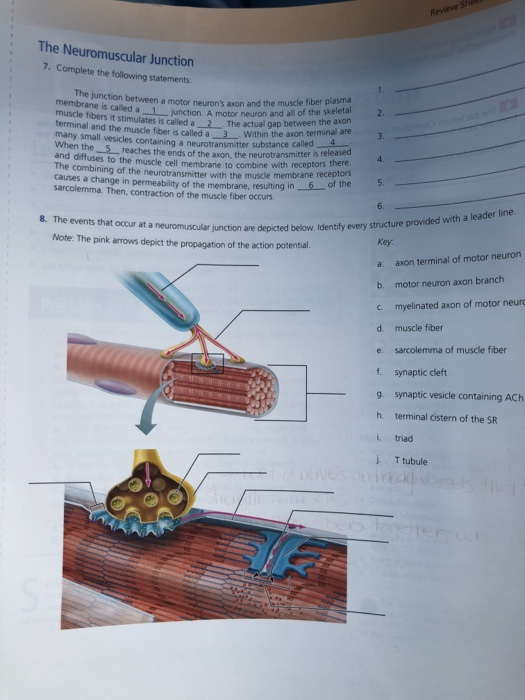
Using The Terms From The Key, Identify Each Structure Indicated By A Leader Line Or Bracket.
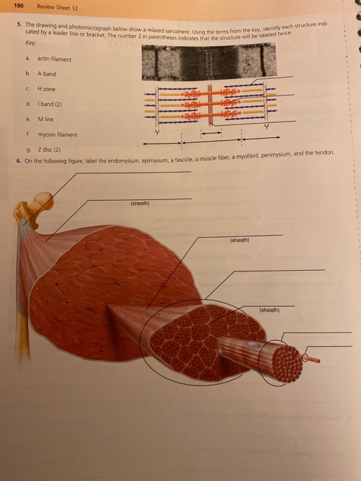
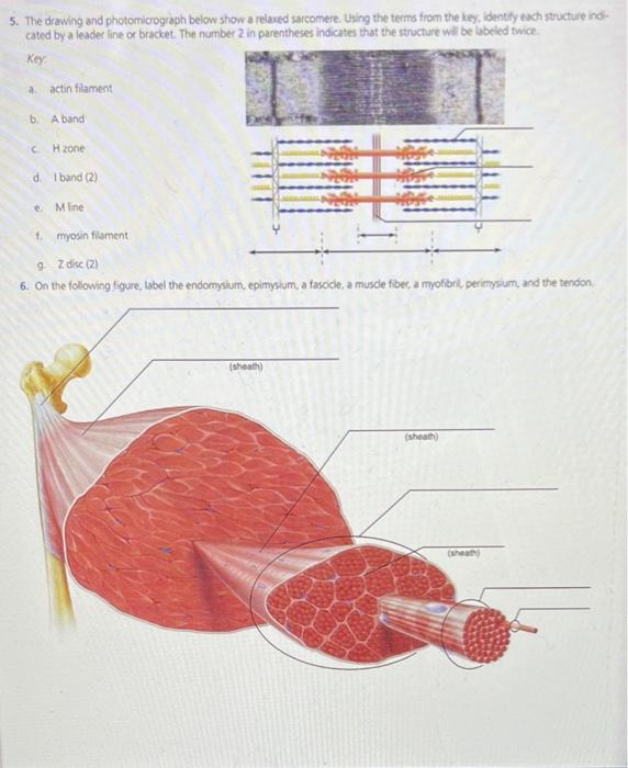
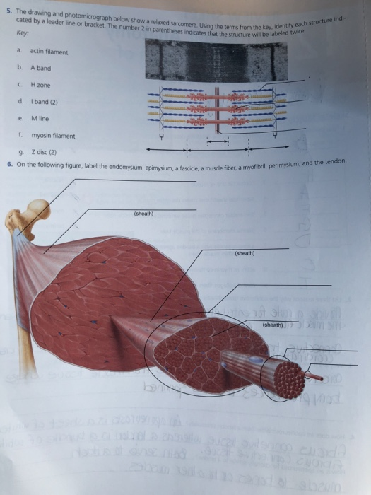
A columnar epithelial cell looks like a column or a tall rectangle. At a basic level, photomicroscopy may be performed simply by connecting a camera to a microscope, thereby enabling the user to take photographs at reasonably high magnification. Using the terms from the key, identity each structure indi cated by a leader ine or bracket. The number 2 in parentheses indicates that the structure will be labeled twice 190 review sheet 12 key:
The Number 2 In Parentheses Indicates That The Structure Will Be Labeled Twice.
The drawing and photomicrograph below show a relaxed sarcomere using the rhe we beldce cated by a leader line or bracket. Details of the structures inside the goblet cell can be seen in an electron micrograph. Each bundle, in turn, is composed of many large peripheral axons. A band c, h zone d.
The Number 2 In Parentheses Indicates That The Structure Will Be Labeled Twice Key A Actin Filament B.
But in differing proportions and with different wall thicknesses. The number 2 in parentheses indicates that the structure will be labeled twice. Using the terms from the key, identify each struc ture indicated by a leader line or bracket. The number 2 in parentheses indicates that the structure will be labeled twice.
A Squamous Epithelial Cell Looks Flat Under A Microscope.
Web the drawing and photomicrograph below show a relaxed sarcomere. Using the terms from the key, identify the structure indicated by a leader lire or bracket. Z disc (2) solution verified answered 6 months ago create a free account to view solutions The number 2 in parentheses indicates that the structure will be labeled twice.