
Spongy Bone Drawing
Spongy Bone Drawing - Center canal or haversian canal of an osteon #5. Web here in this article, you will find a short but essential description of spongy bone with slide images. Compact bone is enclosed, except where it's covered by articular cartilage, and is covered by the periosteum. Internal circumferential lamellae of compact bone #2. Web the bone drawing on the left side of the poster displays the following from outside to inside structure:
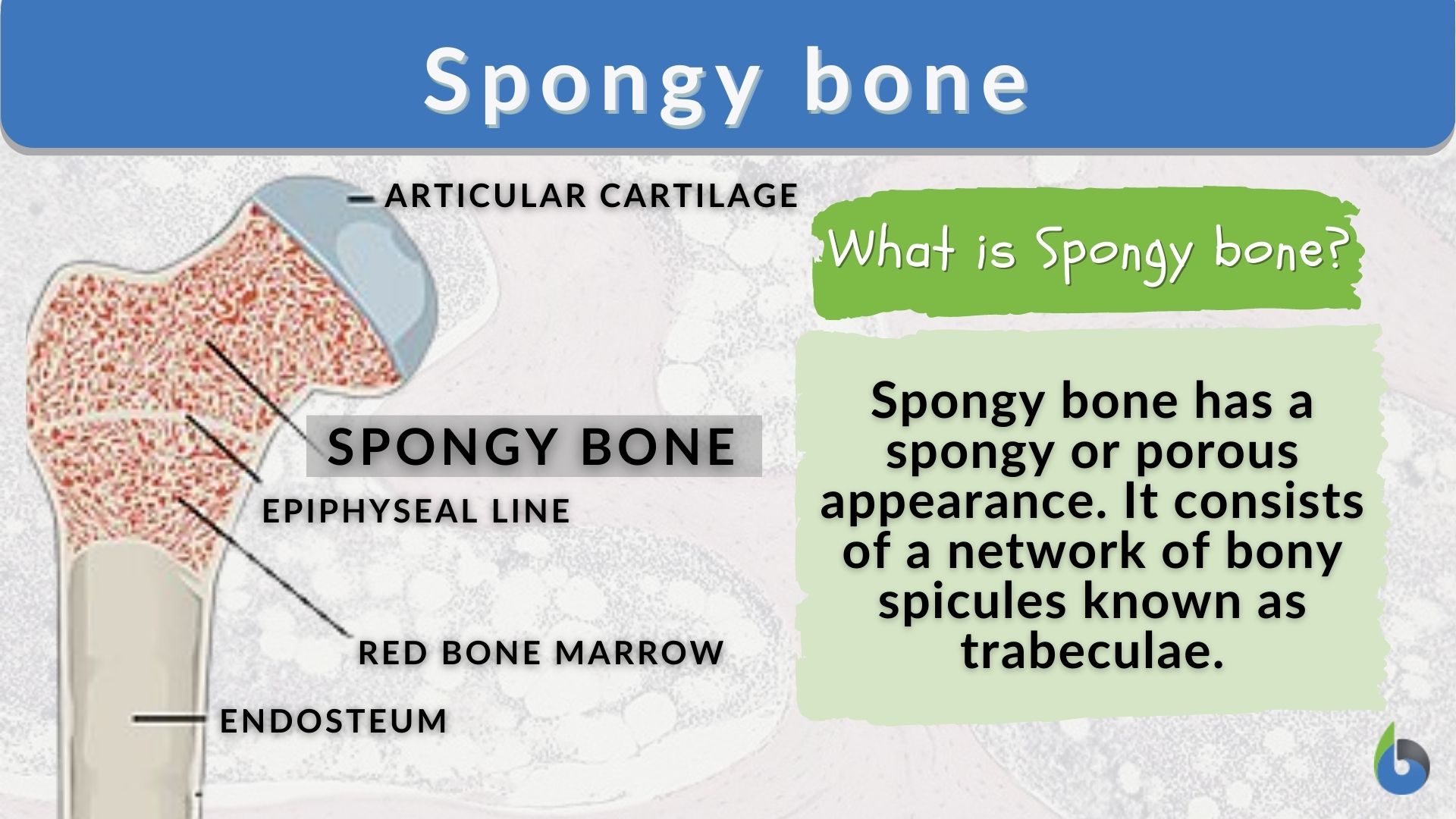
The wider section at each end of the bone is called the epiphysis (plural = epiphyses), which is filled with spongy bone. Describe how bones are nourished and innervated They primarily serve to protect tendons from excess wear. Web here in this article, you will find a short but essential description of spongy bone with slide images. Web spongy bone, also known as cancellous bone or trabecular bone, is a very porous type of bone found in animals. Long, short, flat, sesamoid or irregular. Name five bones of the axial skeleton and five bones of the appendicular skeleton.
Spongy Bone (Cancellous Bone) Definition & Function Biology
The wider section at each end of the bone is called the epiphysis (plural = epiphyses), which is filled with spongy bone. Describe how bones are nourished and innervated They primarily serve to protect tendons from excess wear. Whereas compact bone tissue forms the outer layer of all bones, spongy bone or cancellous bone forms.

Structure of Spongy Bone
Anatomy of a long bone.a typical long bone shows the gross anatomical characteristics of bone. Web basically, in kindergarten when you drew skeletons, you were drawing compact bone. Web about press copyright contact us creators advertise developers terms privacy policy & safety how youtube works test new features nfl sunday ticket press copyright. There are.

Histology of Spongy Bone YouTube
Web structure of bone tissue. Web figure 6.3.1 6.3. Web spongy bone tissue. They primarily serve to protect tendons from excess wear. Web the walls of the diaphysis are composed of dense and hard compact bone. Histology of spongy bone how to draw spongy bone. Web identify the anatomical features of a bone; Ditki is.

Histology Glossary Histology Spongy Bone Draw It to Know It
There are three types of cells that contribute to bone homeostasis. Name five bones of the axial skeleton and five bones of the appendicular skeleton. Web the bone drawing on the left side of the poster displays the following from outside to inside structure: Web the walls of the diaphysis are composed of dense and.

Spongy bone, illustration Stock Image F017/2392 Science Photo Library
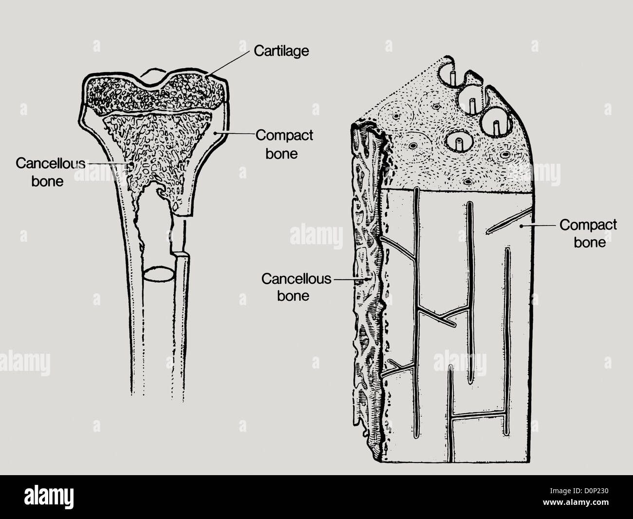
The expanded drawing displays the internal structure. A cross section of the bone shows compact bone and blood vessels in the bone marrow. Web spongy bone tissue. Drawing shows spongy bone, red marrow, and yellow marrow. Web here in this article, you will find a short but essential description of spongy bone with slide images..
Spongy bone Definition and Examples Biology Online Dictionary
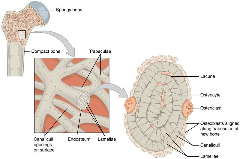
Web like compact bone, spongy bone, also known as cancellous bone, contains osteocytes housed in lacunae, but they are not arranged in concentric circles. Write your answers on the spaces provided. Describe the histology of bone tissue; Lacunae in concentric lamellae of haversian system #7. Spongy bone is usually located at the ends of the.

Spongy Bone Containg Red Bone Marrow Anatomy bones, Basic anatomy and
Diagram illustrating the anatomy of a long bone. Web spongy bone, also known as cancellous bone or trabecular bone, is a very porous type of bone found in animals. Also shown are red blood cells, white blood cells, platelets, and a blood stem cell. It is highly vascularized and contains red bone marrow. Ditki is.

Explanation of bone structure. Two main types of bone include spongy
It is highly vascularized and contains red bone marrow. The wider section at each end of the bone is called the epiphysis (plural = epiphyses), which is filled with spongy bone. Whereas compact bone tissue forms the outer layer of all bones, spongy bone or cancellous bone forms the inner layer of all bones. There.

Healthy Bone Structure Spongy Cancellous Bone. A Bone Is Composed Of
Center canal or haversian canal of an osteon #5. Periosteum, medullary cavity, compact bone, and spongy bone. Drawing shows spongy bone, red marrow, and yellow marrow. Internal circumferential lamellae of compact bone #2. Compare and contrast compact and spongy bone; The names imply that the two types differ in density, or how tightly the tissue.
A line drawing showing the structure in bone, including cancellous or
Web spongy bone, also known as cancellous bone or trabecular bone, is a very porous type of bone found in animals. Center canal or haversian canal of an osteon #5. Web the walls of the diaphysis are composed of dense and hard compact bone. Concentric lamellae of osteon or haversian system #6. This category is.
Spongy Bone Drawing Overall osteon or haversian system of bone #4. External circumferential lamellae of compact bone #3. Concentric lamellae of osteon or haversian system #6. Diagram illustrating the anatomy of a long bone. Inside the compact bone is the periosteum (outer fibrous layer and inner osteogenic layer), interstitial lamellae,.
The Wider Section At Each End Of The Bone Is Called The Epiphysis (Plural = Epiphyses), Which Is Filled With Spongy Bone.
Identify the structures that compose compact and spongy bone; The names imply that the two types differ in density, or how tightly the tissue is packed together. Inside the compact bone is the periosteum (outer fibrous layer and inner osteogenic layer), interstitial lamellae,. There are three types of cells that contribute to bone homeostasis.
Periosteum, Medullary Cavity, Compact Bone, And Spongy Bone.
Concentric lamellae of osteon or haversian system #6. Web spongy bone tissue. They primarily serve to protect tendons from excess wear. So, if you are interested to learn spongy bone histology a then continue this article till the end.
Describe The Histology Of Bone Tissue;
Also shown are red blood cells, white blood cells, platelets, and a blood stem cell. Describe how bones are nourished and innervated Web about press copyright contact us creators advertise developers terms privacy policy & safety how youtube works test new features nfl sunday ticket press copyright. External circumferential lamellae of compact bone #3.
Anatomy Of A Long Bone.a Typical Long Bone Shows The Gross Anatomical Characteristics Of Bone.
Web spongy is a poor description for something that is forming the 'flying buttresses' of our bones. Web like compact bone, spongy bone, also known as cancellous bone, contains osteocytes housed in lacunae, but they are not arranged in concentric circles. You will also find the spongy bone histology slide drawing tutorial at the end of this article. A cross section of the bone shows compact bone and blood vessels in the bone marrow.