Pituitary Gland Drawing
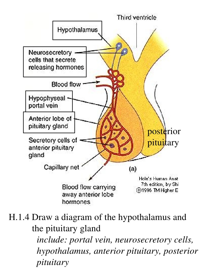
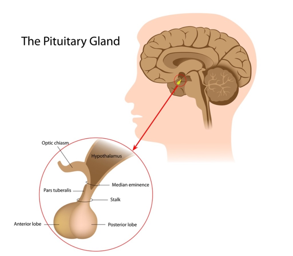
Pituitary Gland Drawing - It is situated in a bony structure called the pituitary fossa, just below the hypothalamus, close to the optic nerve. It consists of two lobes that arise from distinct parts of embryonic tissue: Posterior pituitary the posterior pituitary is actually an extension of the neurons of the paraventricular and supraoptic nuclei of the hypothalamus. The posterior pituitary (neurohypophysis) is neural tissue, whereas the anterior pituitary (also known as the adenohypophysis) is glandular tissue that develops from the primitive. Web drawing the pituitary gland diagram using a samsung stylus.
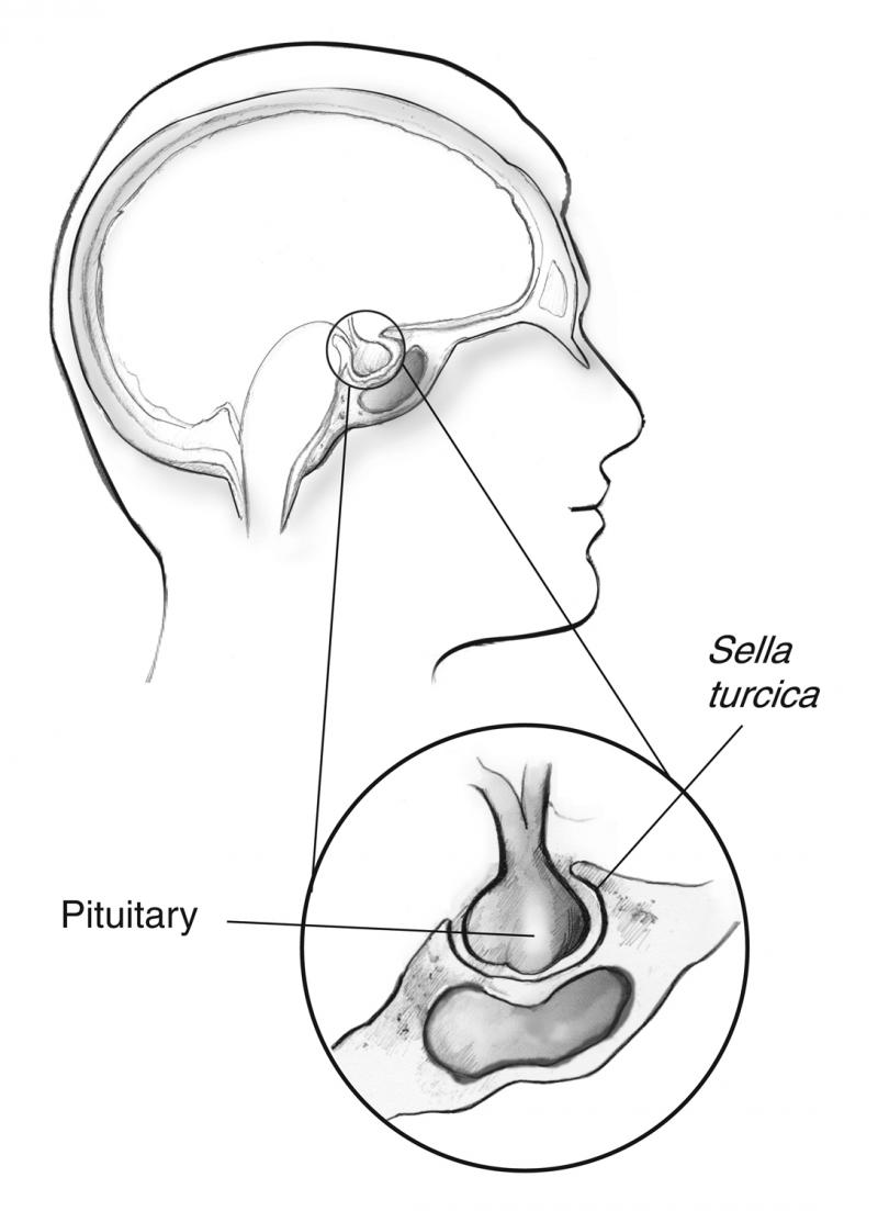
The pituitary gland is a part of your endocrine system. The human pituitary gland is oval shaped, about the size of a chickpea, and weighs 0.5 grams (0.018 oz) on average. Web explanation of histology of pituitary gland with drawing and picture of histology slide | anatomy| explanation | with drawing | histology practical slide / p. Web the pituitary gland is cradled within the sellaturcica of the sphenoid bone of the skull. Give this video a like and subscribe to my channel to watch more! Its main function is to secrete hormones into your bloodstream. The pituitary gland undergoes rapid growth from birth to adult life to reach a weight of 500 mg.
Pituitary Gland Anatomy and Physiology Endocrine System
Posterior pituitary the posterior pituitary is actually an extension of the neurons of the paraventricular and supraoptic nuclei of the hypothalamus. They include benign tumors, several health conditions, pregnancy, and certain medications. It is often referred to as the ‘master gland’ because it produces some of the important hormones in the body. The adult gland.
Pituitary Gland Anatomy, Function, and Treatment
There is a discrepancy between the size of the gland in males and females. The pituitary gland consists of two anatomically and functionally distinct regions, the anterior lobe (adenohypophysis) and the posterior lobe (neurohypophysis). The pituitary region is characterized by an exceptional aggregation of important anatomical structures that serve a unique diversity of essential functions..
Pituitary Gland Drawing at GetDrawings Free download
It consists of two lobes that arise from distinct parts of embryonic tissue: This chapter reviews and illustrates the normal anatomy and mri appearances of the pituitary gland and hypothalamic region. Web the pituitary gland is cradled within the sellaturcica of the sphenoid bone of the skull. Web diagram conditions symptoms health tips what is.
Structure of pituitary gland, illustration Stock Image C039/2020
This gland commonly develops pathology, which may result in a mass effect on adjacent intracranial structures or be hormonally active. Posterior pituitary the posterior pituitary is actually an extension of the neurons of the paraventricular and supraoptic nuclei of the hypothalamus. The anterior pituitary (front lobe) and the posterior pituitary (back lobe). The pituitary gland.
Pituitary Gland The Master Gland
Web introduction the pituitary endocrine gland, which is located in the bony sella turcica, is attached to the base of the brain and has a unique connection with the hypothalamus. Web we provide an overview of the molecular drivers of pituitary organogenesis and illustrate the anatomy and histology of the mature pituitary, comprising adenohypophysis (anterior.
Pituitary Gland Function, Disorders & Pituitary Gland Tumors
Web the pituitary gland (or hypophysis cerebri) is an endocrine gland in vertebrates. In adults, the vertical diameter is approximately 8mm, with the horizontal circumference found to be 12 millimeters (mm). It is often referred to as the ‘master gland’ because it produces some of the important hormones in the body. The pituitary is the.
Pituitary Gland Illustrations, RoyaltyFree Vector Graphics & Clip Art
Posterior pituitary the posterior pituitary is actually an extension of the neurons of the paraventricular and supraoptic nuclei of the hypothalamus. They include benign tumors, several health conditions, pregnancy, and certain medications. The posterior pituitary (neurohypophysis) is neural tissue, whereas the anterior pituitary (also known as the adenohypophysis) is glandular tissue that develops from the.
Pituitary Gland Drawing at GetDrawings Free download
They include benign tumors, several health conditions, pregnancy, and certain medications. The anterior pituitary (front lobe) and the posterior pituitary (back lobe). Web mri is the primary imaging modality for the pituitary gland. It is involved in nearly all processes of homeostasis, as well as growth and development. Web the pituitary gland is anatomically and.
Pituitary Gland Anatomy. Hormones Stock Vector Illustration of head
Web the pituitary gland is cradled within the sellaturcica of the sphenoid bone of the skull. This gland commonly develops pathology, which may result in a mass effect on adjacent intracranial structures or be hormonally active. Web mri is the primary imaging modality for the pituitary gland. The hypothalamus is the pivotal center for the.
Photo of the Pituitary Gland
An enlarged pituitary gland has many potential causes. In humans, the pituitary gland is located at the base of the brain, protruding off the bottom of the hypothalamus. Web we provide an overview of the molecular drivers of pituitary organogenesis and illustrate the anatomy and histology of the mature pituitary, comprising adenohypophysis (anterior lobe), neurohypophysis.
Pituitary Gland Drawing Web the pituitary gland consists of an anterior and posterior lobe, with each lobe secreting different hormones in response to signals from the hypothalamus. In adults, the vertical diameter is approximately 8mm, with the horizontal circumference found to be 12 millimeters (mm). Web drawing the pituitary gland diagram using a samsung stylus. Web the pituitary gland is anatomically and functionally closely related to the hypothalamus. The human pituitary gland is oval shaped, about the size of a chickpea, and weighs 0.5 grams (0.018 oz) on average.
Web Introduction The Pituitary Endocrine Gland, Which Is Located In The Bony Sella Turcica, Is Attached To The Base Of The Brain And Has A Unique Connection With The Hypothalamus.
The pituitary gland undergoes rapid growth from birth to adult life to reach a weight of 500 mg. In humans, the pituitary gland is located at the base of the brain, protruding off the bottom of the hypothalamus. Web gross anatomy of the pituitary gland. Web explanation of histology of pituitary gland with drawing and picture of histology slide | anatomy| explanation | with drawing | histology practical slide / p.
The Posterior Pituitary (Neurohypophysis) Is Neural Tissue, Whereas The Anterior Pituitary (Also Known As The Adenohypophysis) Is Glandular Tissue That Develops From The Primitive.
Drawing shows the hypothalamus, pituitary gland,. An enlarged pituitary gland has many potential causes. The adult gland has an anteroposterior diameter of 8 mm and a transverse diameter of 12 mm. The term hypophysis (from the greek for “lying under”)—another name for the pituitary—refers to the.
In Adults, The Vertical Diameter Is Approximately 8Mm, With The Horizontal Circumference Found To Be 12 Millimeters (Mm).
It is often referred to as the ‘master gland’ because it produces some of the important hormones in the body. Web we provide an overview of the molecular drivers of pituitary organogenesis and illustrate the anatomy and histology of the mature pituitary, comprising adenohypophysis (anterior lobe), neurohypophysis (posterior lobe), pars intermedia and infundibulum (pituitary stalk). Web diagram conditions symptoms health tips what is the pituitary gland? Web your pituitary gland is divided into two main sections:
The Pituitary Region Is Characterized By An Exceptional Aggregation Of Important Anatomical Structures That Serve A Unique Diversity Of Essential Functions.
Give this video a like and subscribe to my channel to watch more! There is a discrepancy between the size of the gland in males and females. They include benign tumors, several health conditions, pregnancy, and certain medications. This chapter reviews and illustrates the normal anatomy and mri appearances of the pituitary gland and hypothalamic region.


/pituitaryanatomyillustration-6c8e132ec19f4e0c92758adfc2a14101.jpg)


