
Microscope Drawing With Label
Microscope Drawing With Label - You can download them individually by clicking the images below, or download them together in a single pdf. Use this with the activity to help students identify and label the main parts of a microscope and then describe their functions. This can be done with pencil and paper or with digital drawing tools. Web establishing a habit of labeling your drawings while discovering how to sketch a microscope slide will enable you to keep your drawing organized. Next, use shades of gray to fill in the eyepiece, illuminator, stage, focus wheels, and lenses.
You can download them individually by clicking the images below, or download them together in a single pdf. First and foremost, we have a labeled microscope diagram, available in both black and white and color. Take a look at your microscope slide and start with the basic shapes and outlines of the objects you see. Label the cell wall, cell membrane, cytoplasm, and chloroplasts in your lab manual. Continue follow my channel and like, share,comment also. There are three structural parts of the microscope i.e. Then, color the base with a light green crayon.
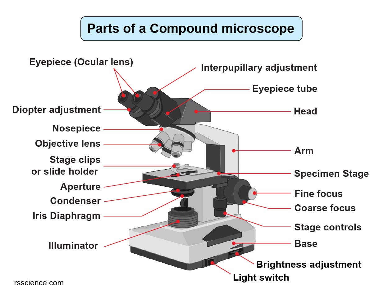
Compound Microscope Parts Labeled Diagram and their Functions Rs
Web 1.1 step 1: Useful as a study guide for learning the anatomy of a microscope. Provide them with diagrams or actual microscopes, and guide them through the function of each part, such as the lens, eyepiece, and stage. Before exploring microscope parts and functions, you should probably understand that the compound light microscope is.
How to Use a Microscope
Web 2.32k subscribers 1k views 3 years ago biology in this video i go over a microscope drawing that is easy with label. It is used to observe things that cannot be seen by the naked eye. Stage and stage clips 7. Outline the slide platform 1.6 step 6: First, color the body of the.

Microscope Drawing And Label at GetDrawings Free download
This short video discuss the expectations of a microscope observation and drawings and also provides examples of errors to watch out for. Web microscope parts and functions with labeled diagram and functions how does a compound microscope work? Shape the microscope head 1.3 step 3: Web parts of the microscope with labeling (also free printouts).

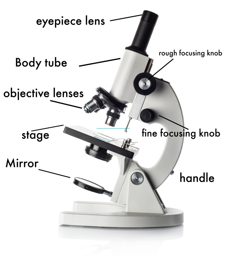
A labeled diagram of a microscope. MLT 101. ) Teaching cells
Also indicate the estimated cell size in micrometers under your drawing. Web a labeled diagram of microscope parts furnishes comprehensive information regarding their composition and spatial arrangement within the microscope, enabling researchers to comprehend their function effectively. Web 1.1 step 1: Shape the illuminator 1.8 step 8: Web parts of the microscope with labeling (also.

Parts of a microscope with functions and labeled diagram
Web sketching the image when you sit down to draw a microscope image, the first step is to sketch out what you see. It is used to observe things that cannot be seen by the naked eye. Web proper microscope drawings and observations. There are three structural parts of the microscope i.e. Web learn about.

Parts of a Compound Microscope Labeled (with diagrams) Medical
Begin with the eyepiece 1.2 step 2: Label the cell wall, cell membrane, cytoplasm, and chloroplasts in your lab manual. Perfect for students or anyone interested in science, this tutorial will guide you. Shape the microscope head 1.3 step 3: Web 1.1 step 1: First and foremost, we have a labeled microscope diagram, available in.

Microscope Diagram to Print 101 Diagrams
Web microscope parts and functions with labeled diagram and functions how does a compound microscope work? Label the cell wall, cell membrane, cytoplasm, and chloroplasts in your lab manual. Knobs (fine and coarse) 6. Diagram of parts of a microscope. Provide them with diagrams or actual microscopes, and guide them through the function of each.

Compound Light Microscope Drawing at GetDrawings Free download
Web in this interactive, you can label the different parts of a microscope. Web in this tutorial, writing master shows you how to draw a realistic microscope with labels step by step. You can download them individually by clicking the images below, or download them together in a single pdf. This activity has been designed.

How to Use a Microscope (Properly) Step by Step New York Microscope
Outline the arm frame 1.4 step 4: This activity has been designed for use in homes and schools. There is a blank copy at the end of the video to review on your own. Draw the base of the microscope sketch 1.7 step 7: Web labeled diagram of a compound microscope major structural parts of.

Microscope diagram Tom Butler Science skills, Microscope parts
Continue follow my channel and like, share,comment also. Before exploring microscope parts and functions, you should probably understand that the compound light microscope is more complicated than just a microscope with more than one lens. Stage and stage clips 7. Web parts of the microscope with labeling (also free printouts) a microscope is one of.
Microscope Drawing With Label Web 1.1 step 1: Web now it’s time to add some color to our drawing of a microscope! Web proper microscope drawings and observations. Web learn about the different parts of the microscope, including the simple microscope and the compound microscope, with labeled pictures and detailed explanations. Draw the objective lenses 1.5 step 5:
It Is Used To Observe Things That Cannot Be Seen By The Naked Eye.
There is a blank copy at the end of the video to review on your own. First, color the body of the microscope with a white crayon. Web in this interactive, you can label the different parts of a microscope. Web learn about the different parts of the microscope, including the simple microscope and the compound microscope, with labeled pictures and detailed explanations.
The Leaf Picture At The Start Of The Article Was Taken Using A Specialized Kind Of Fluorescence Microscopy Called Confocal Microscopy.
Web now it’s time to add some color to our drawing of a microscope! The other type of optical microscope is a. Before exploring microscope parts and functions, you should probably understand that the compound light microscope is more complicated than just a microscope with more than one lens. You can download them individually by clicking the images below, or download them together in a single pdf.
And Drop The Text Labels Onto The Microscope Diagram.
Next, use shades of gray to fill in the eyepiece, illuminator, stage, focus wheels, and lenses. Take a look at your microscope slide and start with the basic shapes and outlines of the objects you see. Each microscope layout (both blank and the version with answers) are available as pdf downloads. Web establishing a habit of labeling your drawings while discovering how to sketch a microscope slide will enable you to keep your drawing organized.
Useful As A Study Guide For Learning The Anatomy Of A Microscope.
This short video discuss the expectations of a microscope observation and drawings and also provides examples of errors to watch out for. Diagram of parts of a microscope. Web 1.1 step 1: Draw the base of the microscope sketch 1.7 step 7: