
Loose Connective Tissue Drawing
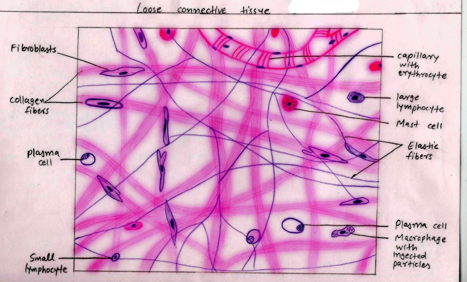
Loose Connective Tissue Drawing - It is the predominant type of connective tissue that joins the cells in the other main tissues (muscle, nerve, and epithelia) and that joins tissues into organs. The drawings of histology images were originally designed to complement the histology component of the first year medical course run prior to 2004. Connective tissue that stores fat and cushions and insulates the body.; As illustrated in figure \(\pageindex{6}\), loose connective tissue has some fibroblasts; Loose connective tissue has some fibroblasts, although macrophages are present as well.
The drawings of histology images were originally designed to complement the histology component of the first year medical course run prior to 2004. Some the epithelial cells are tall and eosinophilic, whereas others are shorter and more basophilic). Web in vertebrates, the most common type of connective tissue is loose connective tissue. You might know the histology of loose connective tissue for further study of different organs or structures from the animals. Web loose irregular connective tissue ; Web #histologydrawing#histology #connectivetissuehistology#connectivetissuediagram#connectivetissue #howtodrawlooseconnectivetissue#howtodrawhistodiagram Web loose connective tissue is the most widely distributed type of connective tissue, found in the lining of the body's inner surfaces.

Histology Drawing of Loose Areolar Tissue with explanation connective
Web the first pages illustrate introductory concepts for those new to microscopy as well as definitions of commonly used histology terms. Web the loose connective tissue is characterized by a loose, irregular arrangement of connective tissue fibers and numerous ground substances. They are sketches from selected slides used in class from the. There are two.

10 b Connective tissue loose areolaradipose... Dense regular
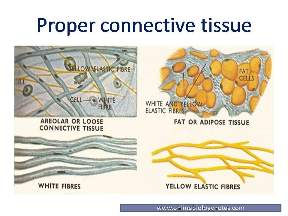
Web connective tissue proper. Web loose connective tissue, also called areolar connective tissue, has a sampling of all of the components of a connective tissue. Connective tissue proper includes both loose (or areolar) connective tissue and dense connective tissue. Connective tissue that stores fat and cushions and insulates the body.; Web the first 6 minutes.

Loose Connective Tissue histology drawingHow to draw loose Connective
It is the predominant type of connective tissue that joins the cells in the other main tissues (muscle, nerve, and epithelia) and that joins tissues into organs. Loose connective tissue, also called areolar connective tissue, has a sampling of all of the components of a connective tissue. Web a connective tissue that is characterised microscopically.

Loose Connective Tissue
Connective tissue proper includes both loose (or areolar) connective tissue and dense connective tissue. Web loose (areolar) connective tissue. Web #histologydrawing#histology #connectivetissuehistology#connectivetissuediagram#connectivetissue #howtodrawlooseconnectivetissue#howtodrawhistodiagram The ecm is composed of a moderate amount of ground substance and two main types of protein fibers: Web about press copyright contact us creators advertise developers terms privacy policy & safety.

Loose Connective Tissue, 40X Histology
Web in drawing images of connective tissue proper preparations seen under the microscope, it is important to simplify the visuals. Dense connective tissues are distinguished microscopically by the close packing of their fibres and relatively few cells and little ground substance. They are sketches from selected slides used in class from the. Web loose connective.
Histology Image Connective tissue
Web loose connective tissue is the most widely distributed type of connective tissue, found in the lining of the body's inner surfaces. The drawings of histology images were originally designed to complement the histology component of the first year medical course run prior to 2004. As illustrated in figure \(\pageindex{6}\), loose connective tissue has some.

What Is Loose Connective Tissue? (preview) Human Anatomy Kenhub
Other components include collagen fibers (c) and elastic fibers (ef) Loose connective tissue has some fibroblasts, although macrophages are present as well. These fibers form an irregular network with spaces between. Its ground substance occupies more volume than the fibers do. Web loose connective tissue is the most widely distributed type of connective tissue, found.
:max_bytes(150000):strip_icc()/loose_connective_tissue-5b68c53446e0fb0050388e5a.jpg)
Connective Tissue Types and Examples
You will find more fibroblasts and macrophages in the sample tissue) #4. Web what are the 3 types of loose connective tissue? Web loose connective tissue, also called areolar connective tissue, has a sampling of all of the components of a connective tissue. Collagen fibers are relatively wide and stain a light pink, while elastic.

20 How to Draw Loose & Dense Regular Connective Tissue/Histology/1st
Web look at the areas outlined in the orientation diagram of the trachea and locate the loose, cellular connective tissue within the glands (the glands are coiled tubes of columnar epithelial cells; Connective tissue preparations are often messy with a number of blotches and shapes irrelevant to the main components of the tissue, which are.
Loose Connective Tissue Reticular
Other components include collagen fibers (c) and elastic fibers (ef) The other specialised types of connective tissue are covered in other topics. Web loose irregular connective tissue ; Web connective tissue | boundless anatomy and physiology histology drawing of loose areolar tissue with explanation | connective. Macrophages are present as well. Connective tissue that stores.
Loose Connective Tissue Drawing There are two types of adipose; As illustrated in figure 1, loose connective tissue has some fibroblasts; Web in vertebrates, the most common type of connective tissue is loose connective tissue. Loose connective tissue, also called areolar connective tissue, has a sampling of all of the components of a connective tissue. Web look at the areas outlined in the orientation diagram of the trachea and locate the loose, cellular connective tissue within the glands (the glands are coiled tubes of columnar epithelial cells;
These Fibers Form An Irregular Network With Spaces Between.
Web loose connective tissue, also known as areolar tissue, is a cellular connective tissue with thin and relatively sparse collagen fibers. Loose connective tissue with nuclei (n) labeled. As illustrated in figure \(\pageindex{6}\), loose connective tissue has some fibroblasts; Loose connective tissue previously included areolar, reticular, and adipose tissues, although this system has been revised to only include areolar tissue.
Web Loose Irregular Connective Tissue ;
Loose connective tissue, also called areolar connective tissue, has a sampling of all of the components of a connective tissue. Its ground substance occupies more volume than the fibers do. The cell to fiber combination makes loose connective tissue flexible but not very resistant to mechanical stress. Macrophages are present as well.
Web Loose Connective Tissue Is The Most Widely Distributed Of All Connective Tissues.
Web loose connective tissue, also called areolar connective tissue, has a sampling of all of the components of a connective tissue. Web connective tissue | boundless anatomy and physiology histology drawing of loose areolar tissue with explanation | connective. It is the predominant type of connective tissue that joins the cells in the other main tissues (muscle, nerve, and epithelia) and that joins tissues into organs. The three types of loose connective tissue include adipose, areolar and basement membrane.
Web In Drawing Images Of Connective Tissue Proper Preparations Seen Under The Microscope, It Is Important To Simplify The Visuals.
You will find more fibroblasts and macrophages in the sample tissue) #4. Dense regular connective tissue which is found in tendons and ligaments, and is shown below. The ecm is composed of a moderate amount of ground substance and two main types of protein fibers: It consists of a loose irregular network of elastin fibers and collagen fibers suspended within a.