
Draw The Structure Of Human Eye And Label Its Parts
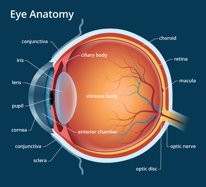
Draw The Structure Of Human Eye And Label Its Parts - Web your eye is a slightly asymmetrical globe, about an inch in diameter. The anatomy of the eye includes auxiliary structures, such as the bony eye socket and extraocular muscles, as well as the structures of the eye itself, such as the lens and the retina. Conjunctiva is the inner layer of the eye. The diagrams show cross sections of the human eyeball; A is the crystalline lens.
Conjunctiva is the inner layer of the eye. Retinal membrane can be imagined as the wall on which the images are projected. The anatomy of the eye includes auxiliary structures, such as the bony eye socket and extraocular muscles, as well as the structures of the eye itself, such as the lens and the retina. As we journey through the different parts, refer to them to better understand their functions. It allows the light to enter our eye to pass through it. Web resource add to collection the human eye contains structures that allow it to perceive light, movement and colour differences. Drag and drop the text labels onto the boxes next to the diagram.
:max_bytes(150000):strip_icc()/eye-conjunctiva-871453538-5a26c6ad22fa3a0037d5edad.jpg)
How the Human Eye Works (Structure and Function)
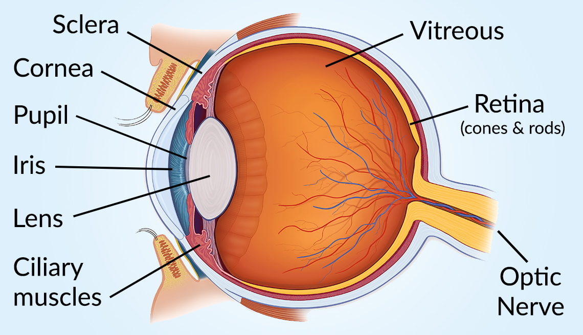
It is a white visible portion. By the end of this activity, students should be able to: The front transparent part of the sclera is called the cornea. Parts of the eye outside the eyeball. It is a non vascular layer. The light passing through cornea, pupil, and lens gets. Web biology biology article diagram.
Human Eye Anatomy, parts and structure Online Biology Notes
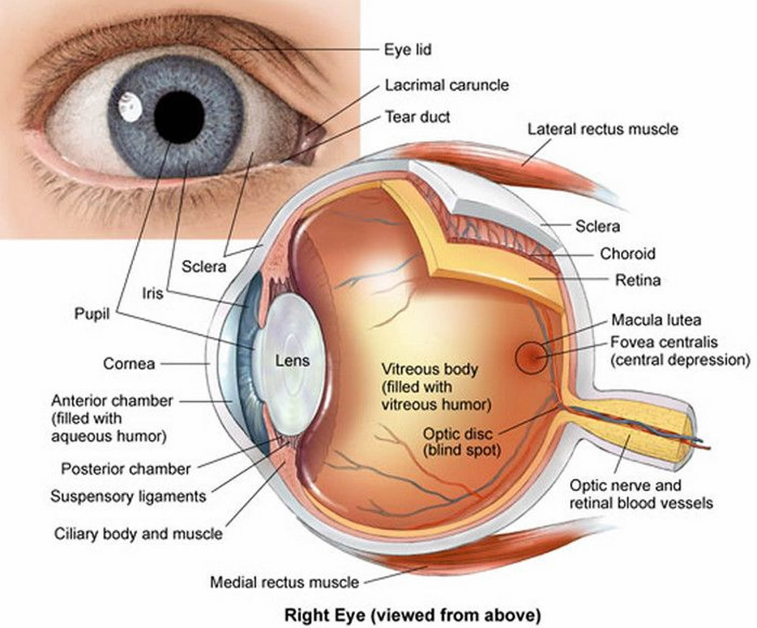
A human eye is roughly 2.3 cm in diameter and is almost a spherical ball filled with some fluid. Here are descriptions of some of the main parts of the eye: It consists of the following parts: A is the crystalline lens. It is a non vascular layer. The eye sits in a protective bony.

OUR EYES WORK LIKE CAMERA’S! Discovery Eye Foundation
The anatomy of the eye includes auxiliary structures, such as the bony eye socket and extraocular muscles, as well as the structures of the eye itself, such as the lens and the retina. As we journey through the different parts, refer to them to better understand their functions. It is the outer covering, a protective.
Human Eye Anatomy Parts of the Eye and Structure of the Human Eye
A human eye is roughly 2.3 cm in diameter and is almost a spherical ball filled with some fluid. Web the external structure of an eye. Choose all answers that apply: The front part (what you see in the mirror) includes: The first thing we're going to draw is the white part of the eye,.
Vision and Eye Diagram How We See
Parts of the eye outside the eyeball. Choose all answers that apply: The diagrams show cross sections of the human eyeball; Conjunctiva is the inner layer of the eye. Working of human eye human eye consists of various parts which help us in seeing the objects, the function of various parts are : Macula lutea.

Labeled Simple Labeled Human Eye Diagram
We will study their structure & func. Web to understand eye problems, it helps to know the different parts that make up the eye and the functions of these parts. Working of human eye human eye consists of various parts which help us in seeing the objects, the function of various parts are : Web.

File1413 Structure of the Eye.jpg Wikimedia Commons
Web your eye is a slightly asymmetrical globe, about an inch in diameter. Macula lutea of the eye. Conjunctiva is the inner layer of the eye. The eye sits in a protective bony socket called the orbit. The size of the human eye can vary, but on average, the adult human eye has a diameter.

Eye diagram by Firkin Human eye diagram, Diagram of the eye, Eye
Web reviewed/revised mar 2022 | modified sep 2022 view professional version the structures and functions of the eyes are complex. Identify the main parts of the human eye Working of human eye human eye consists of various parts which help us in seeing the objects, the function of various parts are : And i'm going.
:max_bytes(150000):strip_icc()/GettyImages-695204442-b9320f82932c49bcac765167b95f4af6.jpg)
Structure and Function of the Human Eye
By the end of this activity, students should be able to: A is the crystalline lens. The cornea is the clear outer part of the eye’s focusing system located at the front of the eye. It consists of the following parts: So i'm just drawing that in. Sclera is the thin white outermost layer of.

Diagram showing the different parts of the eye Parts of the eye, Eye
Choose all answers that apply: Identify the main parts of the human eye And for a description of common vision problems, see refraction and refractive errors: A clear dome over the iris. Web reviewed/revised mar 2022 | modified sep 2022 view professional version the structures and functions of the eyes are complex. Web the structures.
Draw The Structure Of Human Eye And Label Its Parts A thin layer called conjunctiva covers the front portion of the eye. Drag and drop the text labels onto the boxes next to the diagram. Web retina is the innermost layer of the eyeball structure. It is the outer covering, a protective tough white layer called the sclera (white part of the eye). The extraocular muscles are attached to the white part of the eye called the sclera.
Web In This Video, We're Going To Talk About The Structure Of The Eye.
Choose the correct labels for the parts shown. Web structure of human eye. A human eye is roughly 2.3 cm in diameter and is almost a spherical ball filled with some fluid. Web the external structure of an eye.
Web Cornea Of The Eye.
It lines the sclera and is made up of stratified squamous epithelium. Identify the main parts of the human eye It is the outer covering, a protective tough white layer called the sclera (white part of the eye). Web how to draw eye || draw and label the parts of eye.
In This Activity, Students Use Online Or Paper Resources To Identity And Label The Main Parts Of The Human Eye.
It is the transparent membrane which refracts the light entering our eye. By the end of this activity, students should be able to: Iris controls the size of pupil. A is the crystalline lens.
Drag And Drop The Text Labels Onto The Boxes Next To The Diagram.
Web interactive glossary related topics & concepts add to collection + create new collection use this interactive to label different parts of the human eye. And i'm going to label is sclera. The human eye is an organ that detects light and sends signals along the optic nerve to the brain. The most common eye diseases include myopia, hypermetropia, glaucoma and cataract.