
Draw The Digital Slide Of The Esophagus
Draw The Digital Slide Of The Esophagus - Web esophagus histology slide drawing. Examine the small intestine digital slide images. The esophagus is a fibromuscular tube that helps food pass from the mouth through the digestive system. Remember that your drawing should have your name and access code handwritten in the background. Slides 126 and 153 are from the middle 1/3;
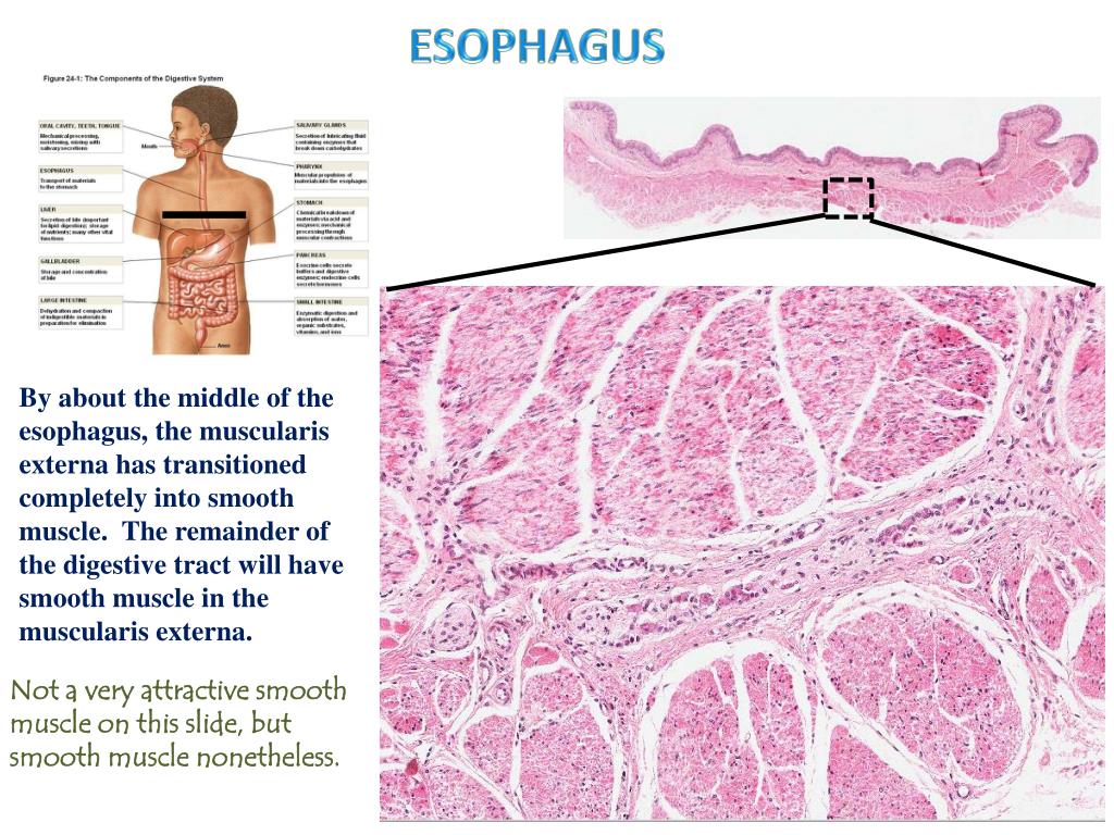
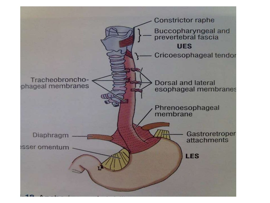
You can easily remove the histology part of the quiz and filter structure types for the quiz yourself. For the purpose of histological descriptions, the esophagus is subdivided into upper (entirely skeletal muscle in the muscularis externa),middle (mixed smooth and skeletal muscle) and lower (entirely smooth muscle) portions. Web this problem has been solved! Be sure to note what structural components of the esophagus are visible. Slides 126 and 153 are from the middle 1/3; Tailor it to your needs! Web a sharp transition in the mucosal epithelium, from stratified squamous moist (esophagus) to simple columnar (cardiac stomach), marks the transition of these two organs.

HISTOLOGY, Epithlium Lab, Esophagus slide
After the food is swallowed, it leaves the mouth and then goes next to the esophagus. Remember that your drawing should have your name and access code handwritten in the background. So, let's focus on that. Remember that your drawing should have your name and access code handwritten in the background. Web esophagus a thick.
PPT Esophagus histology PowerPoint Presentation, free download ID
Do you want to get esophagus histology slide drawing tutorial? The *overall* apparent magnification of a virtual slide is equal to the slide's. When you swallow, food and liquid first move from your mouth to your throat (pharynx). Web the primary function of your esophagus is to carry food and liquid from your mouth to.
anatomy of esophagus by dr ravindra daggupati
A magnification slider control is in the tool bar at the bottom of the microscopic image. Tailor it to your needs! Be sure to note what structural components of the esophagus are visible. Web esophagus histology slide drawing. Esophagus is arranged in 4 concentric layers following a typical gi layering scheme; A small muscular flap.

Esophagus normal histology slides, diagrams, guide (preview) Human
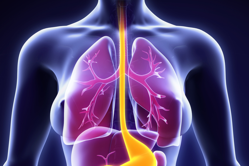
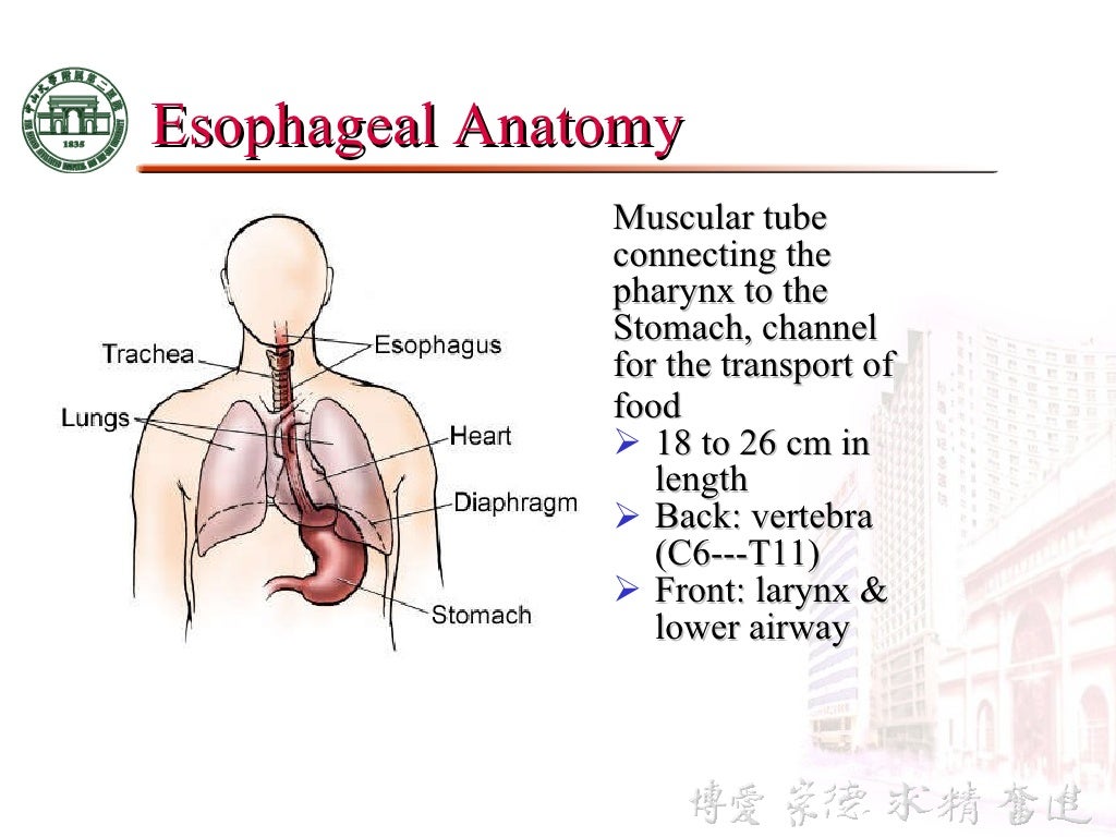
Do you need more images related to esophagus histological. Location of the esophagus in the human body. So, let's focus on that. As described above there are 3 areas of narrowing that occur when. The esophagus can also widen on its own to allow solids to pass through more easily. For the purpose of histological.
Esophagus Facts, Functions & Diseases Live Science
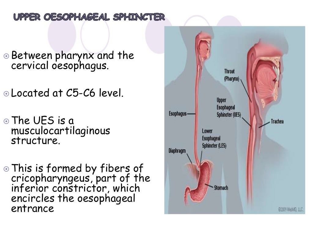
Anatomy and histology start quiz. Remember that your drawing should have your name and access code handwritten in the background. Web watch the complete video on esophagus histology here:. Examine the small intestine digital slide images. A small muscular flap called the epiglottis closes to prevent food and liquid from going down the “ wrong.
PPT Pharynx, Esophagus, Stomach Digital Laboratory PowerPoint
Examine the small intestine digital slide images. The *overall* apparent magnification of a virtual slide is equal to the slide's. That's about as much as two coke bottles, insane! Do you need more images related to esophagus histological. Web esophagus histology slide drawing. The lamina propria underlying the epithelium possesses lymphoid structures and localized mucous.
PPT Esophagus histology PowerPoint Presentation, free download ID
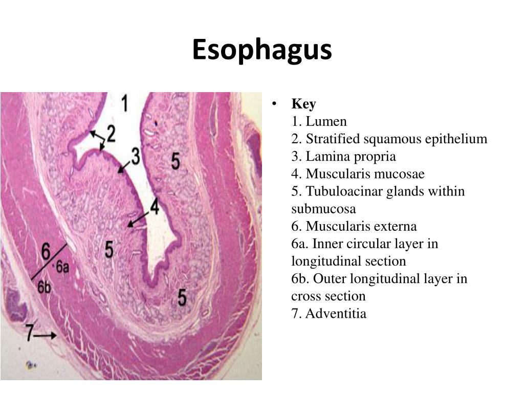
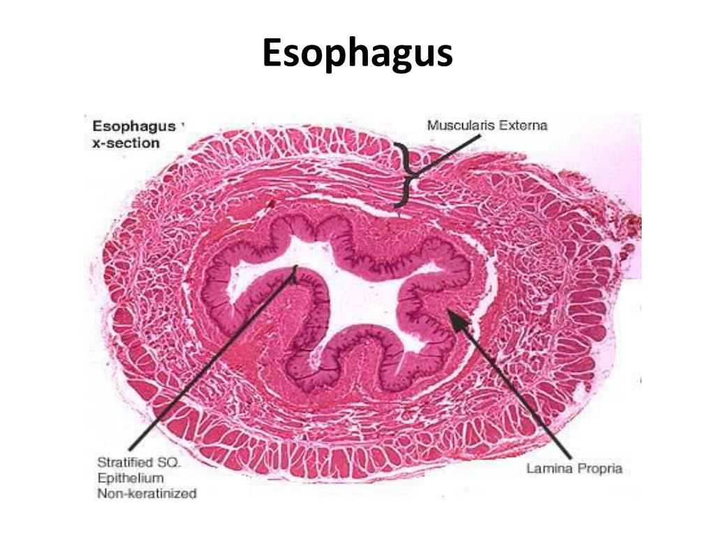
The lamina propria underlying the epithelium possesses lymphoid structures and localized mucous glands (not shown here) in the lower third and sometimes in the upper third. 10x main slide mucosa > submucosa > muscularis externa > adventitia > Do you want to get esophagus histology slide drawing tutorial? And just to make sure, we all.

PPT Esophagus histology PowerPoint Presentation, free download ID
Web esophagus a thick stratified squamous nonkeratinized epithelium lines the esophagus. Web as are all organs opening to the exterior, the esophagus is composed of four tunics: The esophagus is a fibromuscular tube that helps food pass from the mouth through the digestive system. And just to make sure, we all know kind of how.
anatomy of esophagus by dr ravindra daggupati
Additional features of the stomach include the presence of gastric pits extending from the surface to the gastric glands in the lamina propria. And just to make sure, we all know kind of how far the esophagus goes, i'm gonna draw in some lines right here, to show where it starts up here, and then.
3 anatomy & physiology of esophagus
Esophagus is arranged in 4 concentric layers following a typical gi layering scheme; Web test your knowledge on the anatomy and histology of the esophagus and its supplying arteries, veins and nerves in our custom quiz. The esophagus is a fibromuscular tube that helps food pass from the mouth through the digestive system. Web the.
Draw The Digital Slide Of The Esophagus In this question, we are asked to draw the layers of the elementary canal and label them to show the composition and function of each layer, as well as the unique layer which is seen in the stomach, duodenum, and esophagus. The esophagus is a fibromuscular tube that helps food pass from the mouth through the digestive system. Web slide ucsf 226 is from the upper 1/3; Do you want to get esophagus histology slide drawing tutorial? Web the primary function of your esophagus is to carry food and liquid from your mouth to your stomach.
You Can Easily Remove The Histology Part Of The Quiz And Filter Structure Types For The Quiz Yourself.
Location of the esophagus in the human body. So, let's focus on that. Web this problem has been solved! Remember that your drawing should have your name and access code handwritten in the background.
The Esophagus Is A Fibromuscular Tube That Helps Food Pass From The Mouth Through The Digestive System.
After the food is swallowed, it leaves the mouth and then goes next to the esophagus. And adventitia (because the esophagus does not protrude into an internal body cavity). Draw your observations in the space below. Do you need more images related to esophagus histological.
A Small Muscular Flap Called The Epiglottis Closes To Prevent Food And Liquid From Going Down The “ Wrong Pipe ” — Your Windpipe (Trachea).
Web as are all organs opening to the exterior, the esophagus is composed of four tunics: Lab 16 the digestive system draw the digital slide of the esophagus. Do you want to get esophagus histology slide drawing tutorial? Tailor it to your needs!
Use The Hotspot Image Below To Learn More About The Structure And Function Of The Esophagus.
Web watch the complete video on esophagus histology here:. Web the primary function of your esophagus is to carry food and liquid from your mouth to your stomach. Web test your knowledge on the anatomy and histology of the esophagus and its supplying arteries, veins and nerves in our custom quiz. The lamina propria underlying the epithelium possesses lymphoid structures and localized mucous glands (not shown here) in the lower third and sometimes in the upper third.