
Cranial Drawer Sign
Cranial Drawer Sign - In general, radiographic images are used to visualize the instability of the stifle joint by tibial compression, to detect effusion and secondary osteoarthritic changes. Other signs of ccl rupture include “medial buttress” (thickening or scarring on the inside of the knee), and “tibial thrust” (another method to check for cranial displacement of the tibia). The crest is stabilised with a cage and forked tension plate. Web the loss of these normal findings indicates periarticular fibrosis, joint effusion or both. Manual cranial displacement of proximal tibia relative to distal femur ± cranial tibial thrust:
A small bony fragment may be seen at the tibial insertion site of the cranial cruciate ligament in cases of ligament avulsion (22). An examination performed while the patient is sedated is needed to confirm the findings. Web the loss of these normal findings indicates periarticular fibrosis, joint effusion or both. Other signs of ccl rupture include “medial buttress” (thickening or scarring on the inside of the knee), and “tibial thrust” (another method to check for cranial displacement of the tibia). Many patients that do not seem to have a cranial drawer sign while awake have one once they are sedated and relaxed. Often, with a chronic injury, the cranial drawer sign is less effective due some joint stabilization resulting from. Occurs rapidly after ligament rupture in most dogs provides ample indication for surgical joint exploration even in the absence of a.

Cranial Or Anterior Drawer Sign Drawer Gallery
The tta procedure results in a stable stifle joint and eliminates the drawer sign. The veterinarian stabilizes the position of the femur with one hand and. How to perform the cranial drawer test. Most dogs with this injury cannot walk normally and they experience pain. The examiner positions himself by sitting on the examination table.
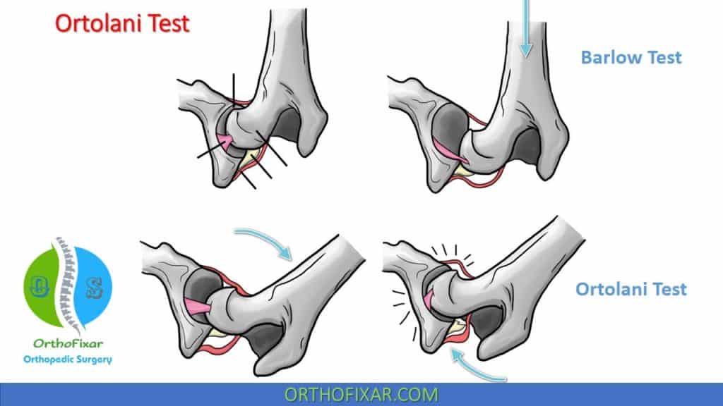
Ortolani Test OrthoFixar 2023
Occurs rapidly after ligament rupture in most dogs provides ample indication for surgical joint exploration even in the absence of a. Web in patients with a partial tear, the cranial drawer sign may or may not be present. Web ± cranial drawer sign: Anesthesia may be necessary to move the limb to the extent needed.
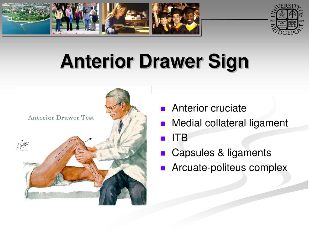
PPT Knee Orthopaedic Tests PowerPoint Presentation, free download
Many patients that do not seem to have a cranial drawer sign while awake have one once they are sedated and relaxed. Web the objective of traditional surgeries, based on the passive model, is the elimination of cranial drawer sign. Web diagnosing cranial cruciate ligament pathology is easy when a supportive history, signalment, gait evaluation,.
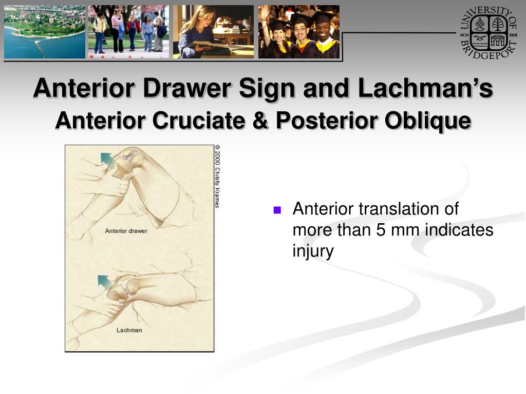
PPT Knee Orthopaedic Tests PowerPoint Presentation, free download
This movement is known as a positive drawer sign. Web patients with chronic ruptures associated with a large amount of scar tissue and arthritis may not exhibit cranial drawer. This test involves manual manipulation of the knee joint and is referred to as the drawer test. Comparing the affected stifle with the normal stifle provides.
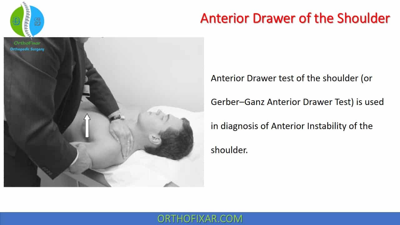
Anterior Drawer Test Shoulder
Occurs rapidly after ligament rupture in most dogs provides ample indication for surgical joint exploration even in the absence of a. Pain upon forced full extension of the stifle is a simple test that is suggestive of early crcld. Web in patients with a partial tear, the cranial drawer sign may or may not be.

Positive cranial drawer sign in a dog with a cranial (anterior
However, some dogs with cranial cruciate ligament pathology do not have palpable stifle instability. Often, with a chronic injury, the cranial drawer sign is less effective due some joint stabilization resulting from. Other signs of ccl rupture include “medial buttress” (thickening or scarring on the inside of the knee), and “tibial thrust” (another method to.
+drawer+test.jpg)
Anterior Drawer Sign Positive Drawer Gallery
Other signs of ccl rupture include “medial buttress” (thickening or scarring on the inside of the knee), and “tibial thrust” (another method to check for cranial displacement of the tibia). Web the loss of these normal findings indicates periarticular fibrosis, joint effusion or both. This movement is known as a positive drawer sign. The examiner.

Cruciate Disease The Cranial Drawer Test YouTube
Other signs of ccl rupture include “medial buttress” (thickening or scarring on the inside of the knee), and “tibial thrust” (another method to check for cranial displacement of the tibia). This test involves manual manipulation of the knee joint and is referred to as the drawer test. Often, with a chronic injury, the cranial drawer.

Drawer Test Bruin Blog
A small bony fragment may be seen at the tibial insertion site of the cranial cruciate ligament in cases of ligament avulsion (22). Web the ability to move the tibia forward (cranially) with respect to a fixed femur is a positive cranial drawer sign indicative of a ccl rupture. The tta procedure results in a.

Drawer Test Bruin Blog
The examiner positions himself by sitting on the examination table in front of the involved knee and grasping the tibia just below the joint line of. Web the loss of these normal findings indicates periarticular fibrosis, joint effusion or both. Cranial movement of tibial tuberosity as hock is manually flexed and gastrocnemius contracts ± meniscal.
Cranial Drawer Sign Occurs rapidly after ligament rupture in most dogs provides ample indication for surgical joint exploration even in the absence of a. Web the ability to move the tibia forward (cranially) with respect to a fixed femur is a positive cranial drawer sign indicative of a ccl rupture. Web is a reliable clinical sign of cruciate rupturepalpation of a medial buttress over the proximal tibia of the affected leg: Web the objective of traditional surgeries, based on the passive model, is the elimination of cranial drawer sign. Web the loss of these normal findings indicates periarticular fibrosis, joint effusion or both.
Occurs Rapidly After Ligament Rupture In Most Dogs Provides Ample Indication For Surgical Joint Exploration Even In The Absence Of A.
Web the loss of these normal findings indicates periarticular fibrosis, joint effusion or both. However, some dogs with cranial cruciate ligament pathology do not have palpable stifle instability. The veterinarian stabilizes the position of the femur with one hand and. Web the key to diagnosis of a ruptured ccl is the demonstration of an abnormal knee motion called the 'cranial drawer sign'.
Cranial Movement Of Tibial Tuberosity As Hock Is Manually Flexed And Gastrocnemius Contracts ± Meniscal Click Ddx:
Web a positive cranial drawer sign can be elicited, and radiographs show joint effusion and cranial dislocation of the tibia (fig. Web a positive tibial compression test and cranial drawer test confirm cclr. Web diagnosing cranial cruciate ligament pathology is easy when a supportive history, signalment, gait evaluation, and radiographic appearance are combined with positive results on tibial compression or cranial drawer tests. How to perform the cranial drawer test.
The Cranial Drawer Test And Tibial Compression Tests Are.
Pain upon forced full extension of the stifle is a simple test that is suggestive of early crcld. Web sliding of the distal femur over the proximal tibia (positive drawer sign) indicates cranial cruciate ligament rupture. In general, radiographic images are used to visualize the instability of the stifle joint by tibial compression, to detect effusion and secondary osteoarthritic changes. Web procedure the patient should be supine with the hips flexed to 45 degrees, the knees flexed to 90 degrees and the feet flat on table.
The Examiner Positions Himself By Sitting On The Examination Table In Front Of The Involved Knee And Grasping The Tibia Just Below The Joint Line Of.
Most dogs with this injury cannot walk normally and they experience pain. Other signs of ccl rupture include “medial buttress” (thickening or scarring on the inside of the knee), and “tibial thrust” (another method to check for cranial displacement of the tibia). An examination performed while the patient is sedated is needed to confirm the findings. Comparing the affected stifle with the normal stifle provides a ready frame of reference.