
Cerebral Cortex Drawing
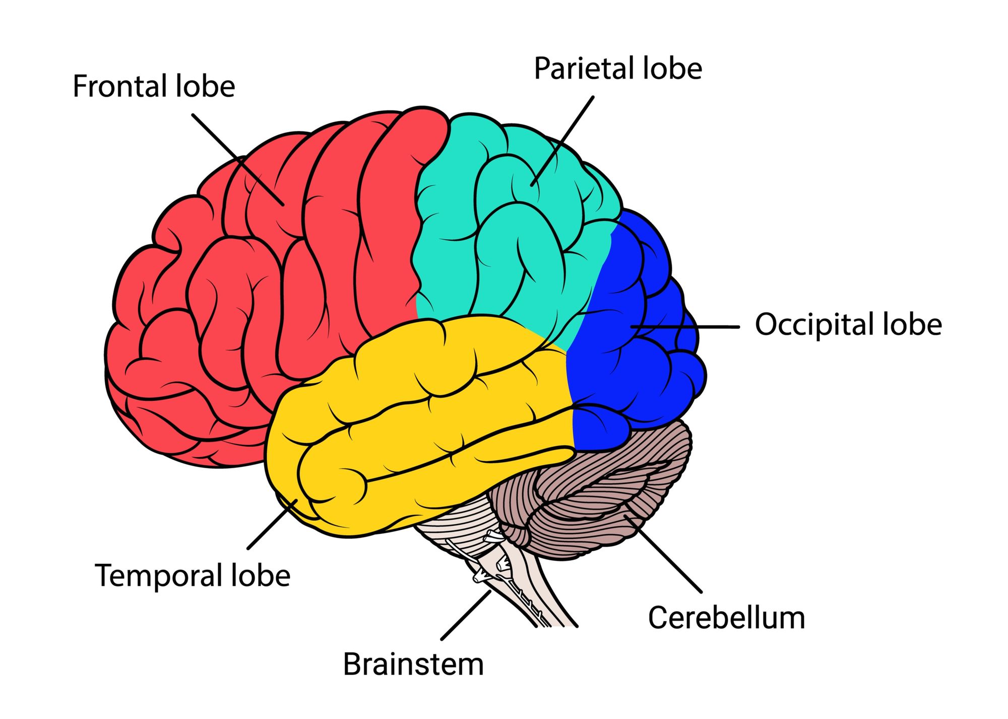
Cerebral Cortex Drawing - The brain, along with the spinal cord, is the main organ of the central nervous system. Frontal, parietal, temporal, and occipital. Structure functions clinical relevance in psychology, the cerebral cortex is defined as the outermost layer of the brain, composed of folded gray matter, playing a. Web the cerebral cortex is the outer covering of the surfaces of the cerebral hemispheres and is folded into peaks called gyri, and grooves called sulci. Between these gyri are grooves or indentations called sulci (singular:
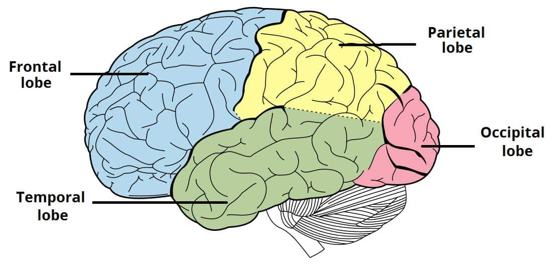
Web the occipital lobe mainly processes vision and the temporal lobe, audition. The cerebral cortex covers the cerebrum and has many folds. It is composed of a series of tortuous folds known as the gyri (singular: The cerebral cortex is 2 to 4 millimeters thick, contains billions of neurons, and has folds that nearly triple its surface area (marieb 2016/p435/c2/para3). “cells in the retina of the eye” (1904), one of. For descriptive purposes each cerebral hemisphere can. Web schematic drawing of six cortical lobes:

Parts Of The Brain Cerebral Cortex Human Anatomy
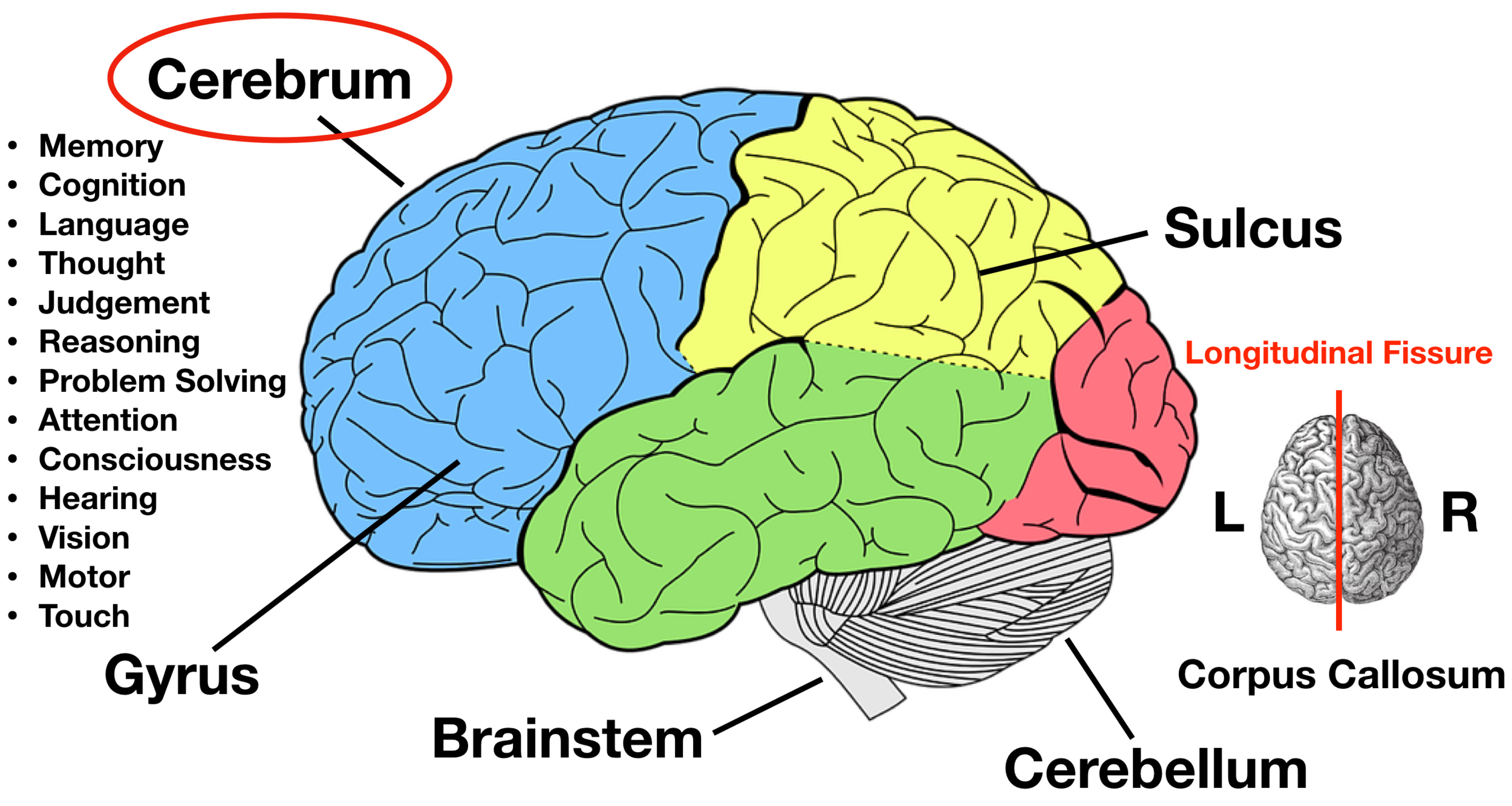
The brain is composed of the cerebrum, cerebellum and brainstem. So the rear of the cerebrum deals with the three main human senses: Anatomical divisions the cerebral cortex is divided in cortical areas based on anatomical location, function and protein expression. External pyramidal layer of cerebral cortex #5. The cerebral cortex covers the cerebrum and.
PostStroke Dizziness How Vestibular Therapy Can Help
The largest part of the brain, the cerebrum initiates and coordinates movement and regulates temperature. It’s two millimeters (mm) to four mm (0.08 inches to 0.16 inches) thick. For descriptive purposes each cerebral hemisphere can. Web learn neuroanatomical highlights of the cerebral cortical areas as an introduction to the clinical highlights of cognitive neurology. This.

Cerebral Cortex Rishi Kathrotia AP Psych 2A
This is a thick area that makes up the largest portion of the brain’s total mass. Anatomical divisions the cerebral cortex is divided in cortical areas based on anatomical location, function and protein expression. It is necessary to keep in mind that these data are old and probably incomplete or erroneous. Web the functional study.
Anatomy human brain areas cerebral cortex with label 1988532 Vector Art
Web the cerebral cortex (cortex of the brain) is the outer grey matter layer that completely covers the surface of the two cerebral hemispheres. Thick, and makes up 40% of the brain's mass. External granular layer of cerebrum structure #4. It’s two millimeters (mm) to four mm (0.08 inches to 0.16 inches) thick. “cells in.

Hand drawn cerebral cortex Vector Free Download
Frontal, parietal, temporal and occipital. The brain is composed of the cerebrum, cerebellum and brainstem. The brain, along with the spinal cord, is the main organ of the central nervous system. This chapter considers the overall organization of cortex from the gross topographic down to the cellular neurophysiology of the cortex. This is a thick.

How to Draw a Brain 14 Steps wikiHow
Internal granular layer of cerebral cortex #6. This chapter considers the overall organization of cortex from the gross topographic down to the cellular neurophysiology of the cortex. The largest part of the brain, the cerebrum initiates and coordinates movement and regulates temperature. The brain, along with the spinal cord, is the main organ of the.
Lobes of the Brain Cerebral Cortex Anatomy, Function, Labeled Diagram
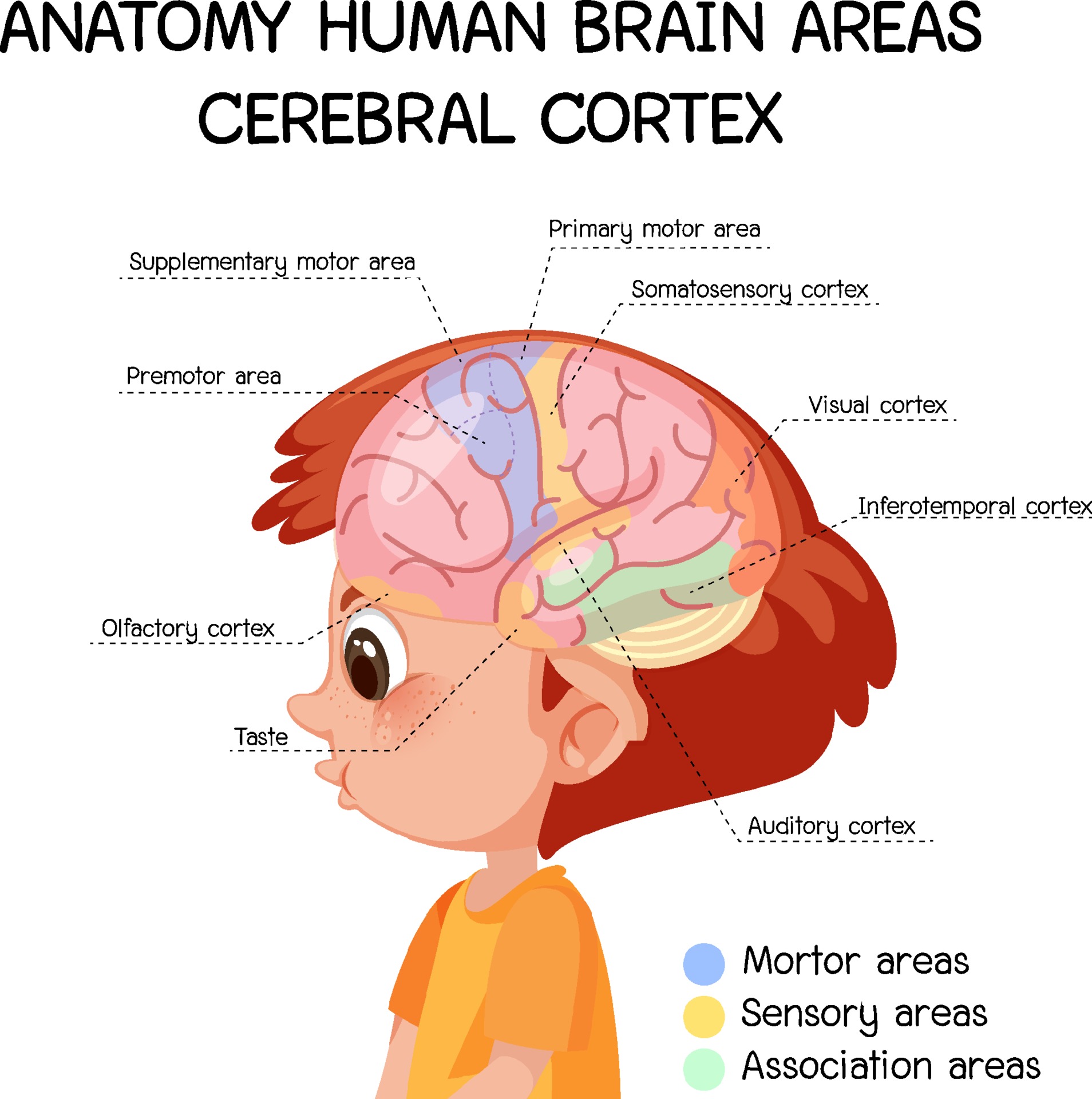
Web the functional study of the cerebral cortex is illustrated by the penfield motor homunculus and brodmann cortical areas, including the broca and wernicke areas. Web cerebrum the cerebrum (front of brain) comprises gray matter (the cerebral cortex) and white matter at its center. Web the area seen from the superolateral view of the cerebrum.
The Brain Concept with Synaesthesia The Syn Moment
Two imaginary lines are drawn on the cerebral hemisphere. Web the cerebral cortex is the largest and most developed part of the human brain and central nervous system (cns). Web the functional study of the cerebral cortex is illustrated by the penfield motor homunculus and brodmann cortical areas, including the broca and wernicke areas. “cells.
Elevated Portions Of The Cerebral Cortex Are Called mapasebab
It is composed of a series of tortuous folds known as the gyri (singular: Web schematic drawing of six cortical lobes: Web the cerebral cortex (cortex of the brain) is the outer grey matter layer that completely covers the surface of the two cerebral hemispheres. Two imaginary lines are drawn on the cerebral hemisphere. The.

Lateral view of the cerebral cortex showing the principal gyri and
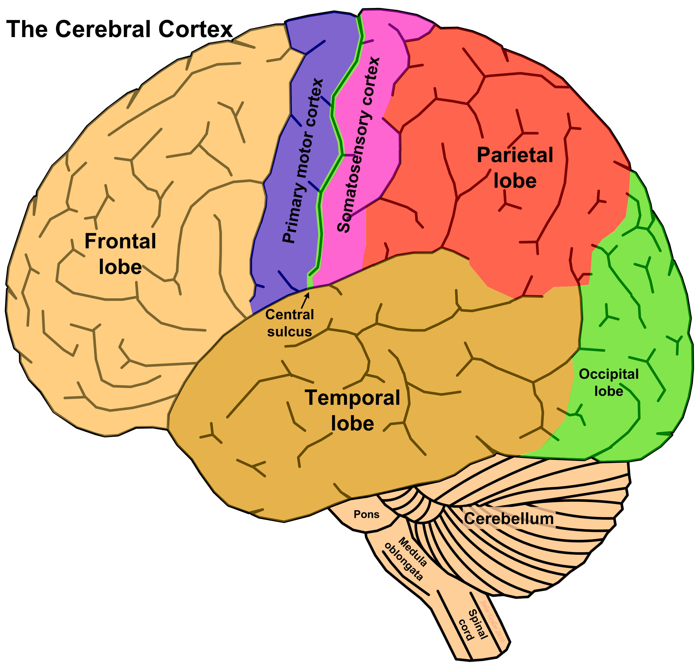
Down the front of the parietal lobe runs a thin strip of somatosensory cortex, which is the term for touch in medical science. Occupying the upper part of the cranial cavity, the cerebral cortex has 4 lobes and is divided into 2 hemispheres that are joined centrally by the corpus callosum. Internal pyramidal layer or.
Cerebral Cortex Drawing Internal pyramidal layer or ganglionic layer of cerebral cortex #7. It is composed of a series of tortuous folds known as the gyri (singular: This outermost layer of the brain integrates information. Occupying the upper part of the cranial cavity, the cerebral cortex has 4 lobes and is divided into 2 hemispheres that are joined centrally by the corpus callosum. External molecular layer of cerebrum #3.
Thick, And Makes Up 40% Of The Brain's Mass.
Web the occipital lobe mainly processes vision and the temporal lobe, audition. It is composed of a series of tortuous folds known as the gyri (singular: Web the cerebral cortex represents the highest level of function of the human brain. Occupying the upper part of the cranial cavity, the cerebral cortex has 4 lobes and is divided into 2 hemispheres that are joined centrally by the corpus callosum.
Your Cortex Is Divided Into Four Lobes:
This layer is thrown into complex folds, with elevations called gyri and grooves known as sulci. It is the most complex organ of the body, with many layers and components that play their roles in almost every function performed by the body. The brain, along with the spinal cord, is the main organ of the central nervous system. Internal pyramidal layer or ganglionic layer of cerebral cortex #7.
So The Rear Of The Cerebrum Deals With The Three Main Human Senses:
The brain is composed of the cerebrum, cerebellum and brainstem. Web the functional study of the cerebral cortex is illustrated by the penfield motor homunculus and brodmann cortical areas, including the broca and wernicke areas. This outermost layer of the brain integrates information. External granular layer of cerebrum structure #4.
The Folds On The Surfaces Of The Cerebral Hemispheres Are Comprised Of Ridges Of Tissue, Called Gyri, Separated By Shallow Grooves, Called Sulci (Marieb 2016/P435/C1/Para 3)
Cognitive functions include thinking, perceiving, and understanding language. It is about 2 to 4 mm thick and contains an aggregation of nerve cell bodies. Web in summary, the cerebral cortex is divided into four lobes that are responsible for processing and interpreting input from various sources and maintaining cognitive function. Web the cerebral cortex (cortex of the brain) is the outer grey matter layer that completely covers the surface of the two cerebral hemispheres.