
Cell Drawing With Labels
Cell Drawing With Labels - Web label lines should be kept to one side of the drawing (in parallel to the top of the page) and drawn with a ruler drawings of cells are typically made when visualizing cells at a higher magnification power, whereas plan drawings are typically made of tissues viewed under lower magnifications (individual cells are never drawn in a plan diagram) A labeled diagram of an animal cell, and a glossary of animal cell terms. Lines are clear and not smudged. Web 7,261 5,292 let us have a detailed look at the plant cell, its structure, and the functions of different plant cell organelles. Web use this convenient study aid in preparation for your upcoming test or quiz.
Be sure to indicate the magnification used and specimen name. Avoid ‘feathery’ pencil lines and gaps. The outermost part of the cell, which is shown as a thick outline of the figure, is labeled cell wall. Include descriptions of what each organelle does. This membrane has about the consistency of.salad oil 1. There are almost no erasures or stray marks on the paper. Download a free printable outline of this video and draw along with us:.

anatomy of the cells
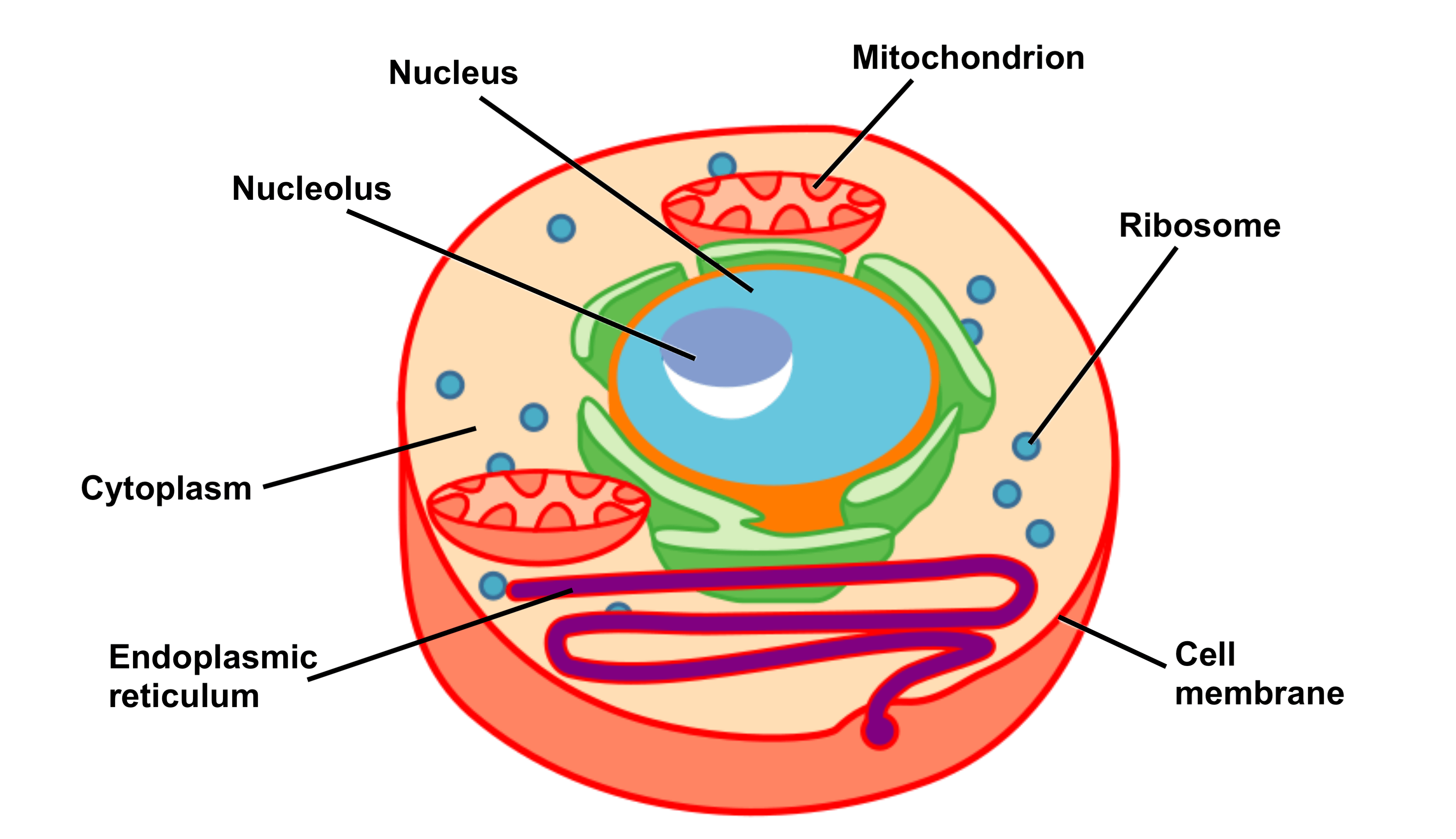
Introduction each cell of your body is encased in a tiny bubble of membrane. Also indicate the estimated cell size in micrometers under your drawing. Using arrows and textables, label parts of a cell and describe each part's function. The cell organelles are enclosed by the plasma membrane including the cell nucleus. Eukaryotic cells are.
Biology Club Our cells 1 ( structure, function, division, disorder
Use a ruler to draw straight, horizontal lines. The labels should form a vertical list. Label one cell with structures listed above. Web a labeled diagram of the animal cell and its organelles. These printables a free for subscribing members of tim’s printables. Where, prokaryotes are just bacteria and archaea, eukaryotes are literally everything else..

Animal Cell Parts Easy Drawing / Draw It Neat How To Draw Animal Cell
The outermost part of the cell, which is shown as a thick outline of the figure, is labeled cell wall. Learn about the different parts of a cell. In the context of the cell cycle, mitosis is the part of the division process in which the dna of the cell's nucleus is split into two.
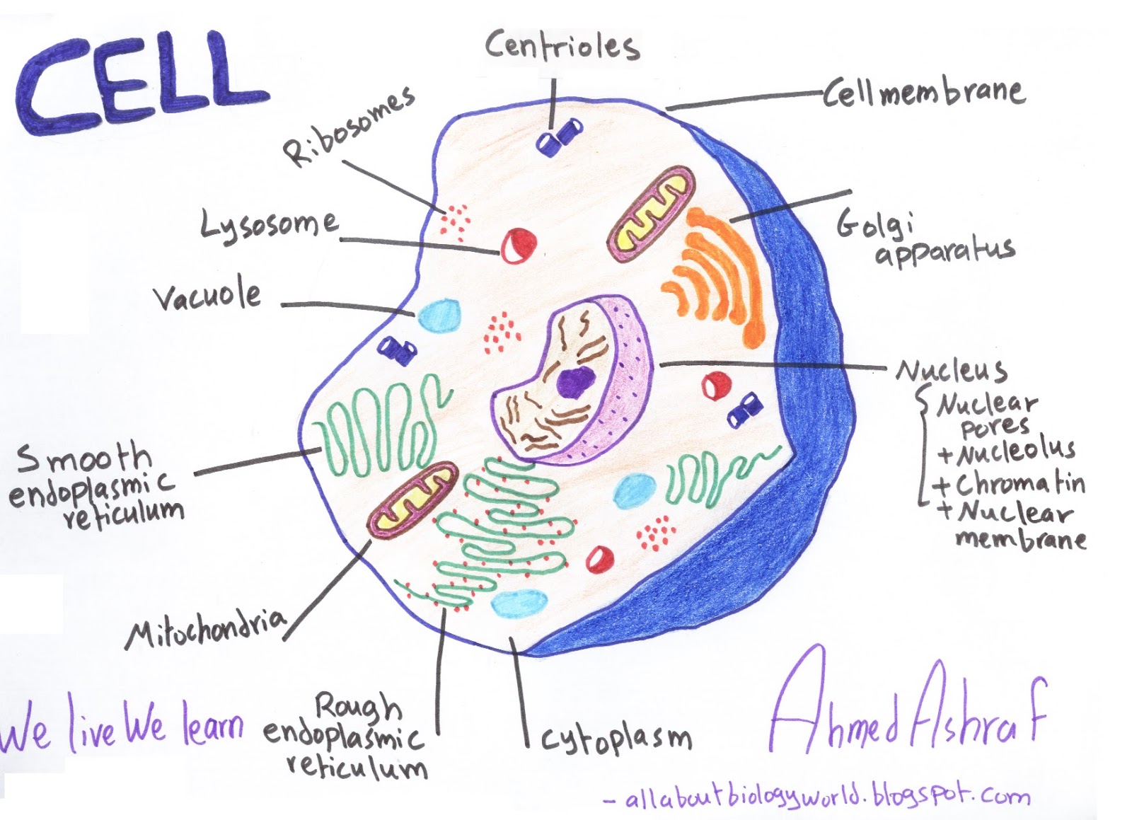
Animal Cell Structure Carlson Stock Art
High power drawings) that you must know! Web mitosis is a type of cell division in which one cell (the mother) divides to produce two new cells (the daughters) that are genetically identical to itself. What purpose do epithelial cells serve? Where, prokaryotes are just bacteria and archaea, eukaryotes are literally everything else. Web complex.
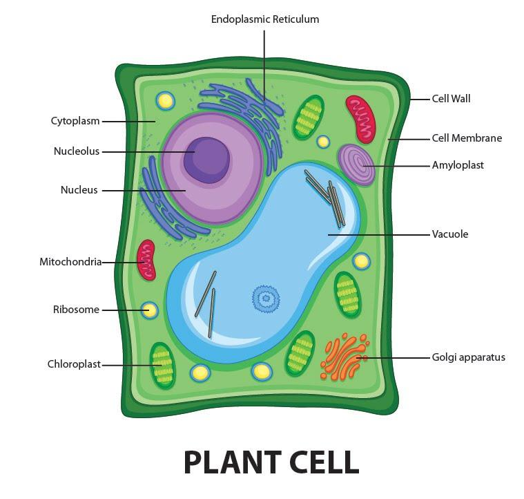
Draw a welllabelled diagram of a plant cell.
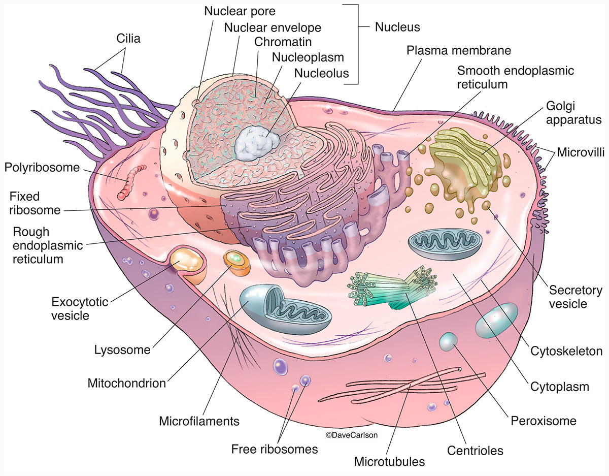
Web find the cell membrane, nucleus, nuclear envelope, and cytoplasm. In the context of the cell cycle, mitosis is the part of the division process in which the dna of the cell's nucleus is split into two equal sets of chromosomes. The cell structure illustrations for these diagrams were generated in biorender. Web cell organelles.

Cells
Lines are clear and not smudged. Be sure to indicate the magnification used and specimen name. Avoid ‘feathery’ pencil lines and gaps. There are six animal cell diagrams to choose from. Color is used carefully to enhance the drawing. Also indicate the estimated cell size in micrometers under your drawing. Web label lines should be.
Cell Structure and Function Part 1 The Organelles Medical Exam Prep
Most cells do not have lysosomes or. Using arrows and textables, label parts of a cell and describe each part's function. There are six animal cell diagrams to choose from. Also, draw an outer circle with a darker shade. Scientists use microscopes to estimate the appearance of structures within the cell and then artists attempt.

What is a cell? Facts
There are almost no erasures or stray marks on the paper. Lines are clear and not smudged. Protein, lipid, and carbohydrate components of the membrane. Web label lines should be kept to one side of the drawing (in parallel to the top of the page) and drawn with a ruler drawings of cells are typically.

South Pontotoc Biology Plant and Animal Cell Diagrams
Create a cell diagram with each part of plant and animal cells labeled. A labeled diagram of an animal cell, and a glossary of animal cell terms. A thinner layer just inside the cell wall is labeled cell membrane. There are various cell organelles, out of which, some are common in most types of cells.

plant cell drawing with labels
This figure show the major organelles and other cell components of a typical eukaryotic plant cell. Introduction each cell of your body is encased in a tiny bubble of membrane. Web label the plant cell diagram using the glossary of plant cell terms. Scientists use microscopes to estimate the appearance of structures within the cell.
Cell Drawing With Labels Draw three representative cells, each about 2 cm in diameter. The cell structure illustrations for these diagrams were generated in biorender. Web you can easily make a human cell drawing with this tutorial. Include descriptions of what each organelle does. Find diagrams of a plant and an animal cell in the science tab.
These H5P Resources Are Made Available Openly With The Cc By License.
Use a ruler to draw straight, horizontal lines. Also, draw an outer circle with a darker shade. The labels should form a vertical list. Web cell structure and function > plasma membranes structure of the plasma membrane google classroom the fluid mosaic model of the plasma membrane.
A Thinner Layer Just Inside The Cell Wall Is Labeled Cell Membrane.
Find diagrams of a plant and an animal cell in the science tab. The outermost part of the cell, which is shown as a thick outline of the figure, is labeled cell wall. Include descriptions of what each organelle does. Avoid ‘feathery’ pencil lines and gaps.
Scientists Use Microscopes To Estimate The Appearance Of Structures Within The Cell And Then Artists Attempt To Create An Interesting Visual Of These.
Label the cell wall, cell membrane, cytoplasm, and chloroplasts in your lab manual. These printables a free for subscribing members of tim’s printables. Protein, lipid, and carbohydrate components of the membrane. This has necessitated some basic changes to how these types of handouts are shared with students.
Color Is Used Carefully To Enhance The Drawing.
A labeled diagram of an animal cell, and a glossary of animal cell terms. The cell structure illustrations for these diagrams were generated in biorender. Web label lines should be kept to one side of the drawing (in parallel to the top of the page) and drawn with a ruler drawings of cells are typically made when visualizing cells at a higher magnification power, whereas plan drawings are typically made of tissues viewed under lower magnifications (individual cells are never drawn in a plan diagram) Structure, parts, functions, labeled diagram june 6, 2023 by faith mokobi edited by: