
Areolar Tissue Drawing
Areolar Tissue Drawing - This video will be very helpful for students to. Web areolar tissue is a type of loose connective tissue found throughout the body. Web areolar connective tissue diagram. For example, it creates telae, such as the tela submucosa and tela subserosa, which attach mucous and serous membranes to the muscular layer. This is a well labelled diagr.
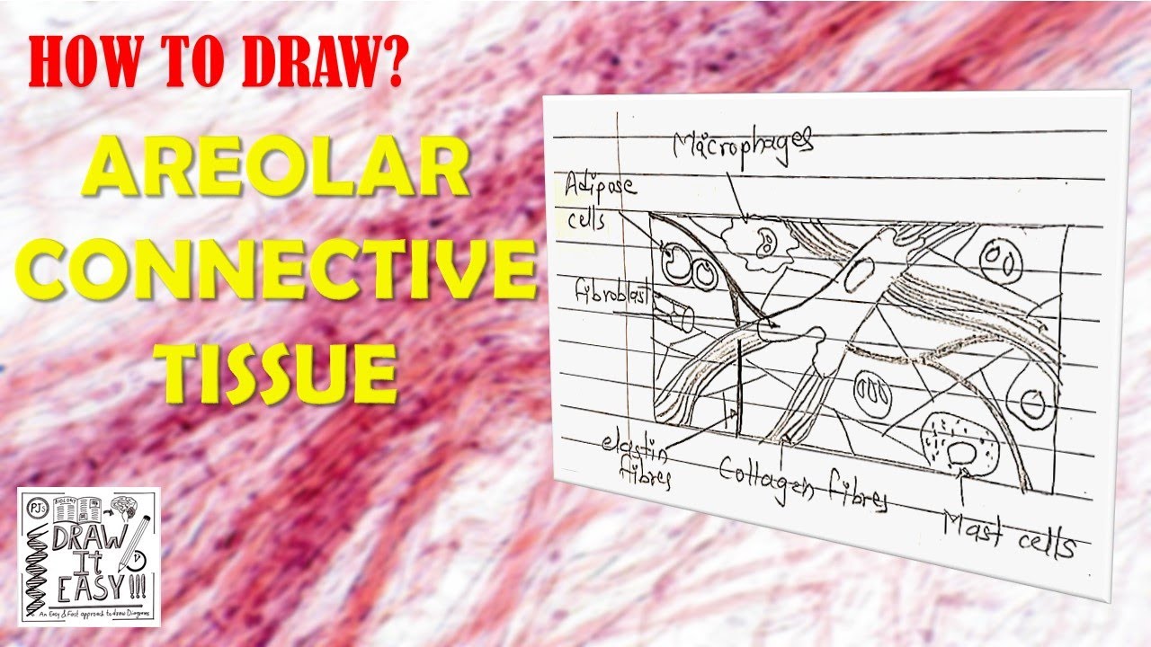
In the circle below, draw a representative sample of key features you identified, taking care to correctly and clearly draw their true shapes and directions. In summary, areolar tissue is tough, yet flexible, and comprises membranes. Web how to draw a diagram of areolar tissue in exam is the topic. Web areolar connective tissue is the most familiar type of connective tissue in vertebrates. Packing material for blood vessels and nerves, dermis of skin, and mucous membranes. Class 9 biology (india) > unit 2. The areolar tissue fills the spaces between the different organs and connects the skin to the underlying muscles.

S19 Educators Science I Class 9 I Chapter 6 I Tissues
In the circle below, draw a representative sample of key features you identified, taking care to correctly and clearly draw their true shapes and directions. Web histology diagrams.loose areolar connective tissue. Web obtain a slide of connective tissue proper/areolar ct from the slide box. Areolar tissue is found beneath the skin (subcutaneous tissue) and surrounds.

Areolar Connective Tissue
View the slide on an appropriate objective. Web q1 what is areolar connective tissue? This is the most abundant tissue in the body, it covers organs, holds blood vessels and nerves in place, forms the dermis of the skin, and the connective tissue layer of mucous membranes. For example, it creates telae, such as the.

Histology Drawing of Loose Areolar Tissue with explanation connective
Class 9 biology (india) > unit 2. Areolar connective tissue has no obvious structure, like layers or rows of cells. Packing material for blood vessels and nerves, dermis of skin, and mucous membranes. Web draw it video tutorial: In the bone marrow as well as around the blood vessels and nerves. Web in summary, areolar.

draw a labeled diagram of areolar tissue Brainly.in
The fibers and other components of the connective tissue matrix are secreted by fibroblasts. Areolar connective tissue has no obvious structure, like layers or rows of cells. Web the areolar tissue is discover under the epidermis layer and is likewise beneath the epithelial cells of all the body plans with exterior openings. Areolar tissue is.
How to Draw Areolar Connective Tissue Biology Diagrams Guide Class
Beneath the dermis lies the hypodermis, which is composed mainly of loose connective. Areolar tissue is found beneath the skin (subcutaneous tissue) and surrounds organs, nerves, blood vessels, and muscle fibers, providing a flexible and resilient support system. It carries organs in place and attaches epithelial tissue to other underlying tissues. Web histology diagrams.loose areolar.

Areolar Connective Tissue
Web areolar connective tissue diagram. Areolar tissue is the least specialized type of connective tissue proper with a matrix containing interwoven yet loosely arranged (widely spaced) elastic and collagen fibers in a thick ground substance that fills most of. Fill out the blanks next to your drawing. No views 1 minute ago. Areolar connective tissue.

How to draw areolar tissue most easy way YouTube
Fill out the blanks next to your drawing. It is likewise a component of the lamina propria of the gastrointestinal and respiratory tracts. Beneath the dermis lies the hypodermis, which is composed mainly of loose connective. Web q1 what is areolar connective tissue? Areolar connective tissue has no obvious structure, like layers or rows of.

areolar tissue 40X labeled fibroblasts Google Search anatomy
This is a well labelled diagr. The areolar tissue is a loose connective tissue that can be seen between the skin and muscles; As stated earlier, the areolar tissue is the. Web figure 5.2 layers of skin the skin is composed of two main layers: Fill out the blanks next to your drawing. Description of.

Areolar Connective Tissue Diagram Quizlet
Areolar connective tissue has no obvious structure, like layers or rows of cells. Web obtain a slide of connective tissue proper/areolar ct from the slide box. This is the well labelled diagram of structure of areolar tissue. This video will be very helpful for students to. In the circle below, draw a representative sample of.

draw a labeled diagram of areolar tissue Brainly.in
Muscular tissue and neural tissue. Web obtain a slide of connective tissue proper/areolar ct from the slide box. Areolar connective tissue has no obvious structure, like layers or rows of cells. In the circle below, draw a representative sample of key features you identified, taking care to correctly and clearly draw their true shapes and.
Areolar Tissue Drawing It carries organs in place and attaches epithelial tissue to other underlying tissues. Packing material for blood vessels and nerves, dermis of skin, and mucous membranes. This video will be very helpful for students to. Web figure 5.2 layers of skin the skin is composed of two main layers: Web areolar connective tissue is the most familiar type of connective tissue in vertebrates.
View The Slide On An Appropriate Objective.
Web 29.4k subscribers subscribe 4.8k views 2 years ago #diagram #biology #asapknowledge hello friends, this is my youtube channel and in this channel i used to share videos of different diagrams in. In the bone marrow as well as around the blood vessels and nerves. Web histology diagrams.loose areolar connective tissue. Web in summary, areolar tissue is tough, yet flexible, and comprises membranes.
In Summary, Areolar Tissue Is Tough, Yet Flexible, And Comprises Membranes.
As stated earlier, the areolar tissue is the. This is a well labelled diagr. Web loose connective tissue (lct), also called areolar tissue, belongs to the category of connective tissue proper. Beneath the dermis lies the hypodermis, which is composed mainly of loose connective.
Areolar Tissue Is The Least Specialized Type Of Connective Tissue Proper With A Matrix Containing Interwoven Yet Loosely Arranged (Widely Spaced) Elastic And Collagen Fibers In A Thick Ground Substance That Fills Most Of.
The areolar tissue is a loose connective tissue that can be seen between the skin and muscles; Areolar connective tissue has no obvious structure, like layers or rows of cells. It is characterised by its loose arrangement of cells and abundant extracellular matrix. Web loose connective tissue is found around every blood vessel and helps to keep the vessel in place.
Web How To Draw A Diagram Of Areolar Tissue In Exam Is The Topic.
Web figure 5.2 layers of skin the skin is composed of two main layers: This is the most abundant tissue in the body, it covers organs, holds blood vessels and nerves in place, forms the dermis of the skin, and the connective tissue layer of mucous membranes. This is the well labelled diagram of structure of areolar tissue. Its cellular content is highly abundant and varied.